Category:Cerebellar vermis
anatomical structure in the brain  | |||||
| Upload media | |||||
| Instance of |
| ||||
|---|---|---|---|---|---|
| Subclass of |
| ||||
| |||||
Media in category "Cerebellar vermis"
The following 59 files are in this category, out of 59 total.
-
 1613 Major Regions of the Cerebellum-02.jpg 2,248 × 1,167; 976 KB
-
Cerebellar hemisphere by Sanjoy Sanyal.webmsd.webm 45 s, 718 × 404; 4.63 MB
-
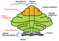
 Cerebellar lobes and lobules.png 1,961 × 1,672; 611 KB
-
 Cerebellar vermis - 03.png 1,200 × 1,200; 982 KB
-
 Cerebellar vermis - 06.png 1,200 × 1,200; 1.02 MB
-
 Cerebellar vermis - animation.gif 600 × 600; 8.55 MB
-
 Cerebellar vermis -- 01.png 1,200 × 1,200; 964 KB
-
 Cerebellar vermis -- 03.png 1,200 × 1,200; 951 KB
-
 Cerebellar vermis -- 04.png 1,200 × 1,200; 1.08 MB
-
 Cerebellar vermis -- 05.png 1,200 × 1,200; 998 KB
-
 Cerebellar vermis -- 06.png 1,200 × 1,200; 1,021 KB
-
 Cerebellar vermis -- animation.gif 600 × 600; 6.78 MB
-
 Cerebellar vermis --- 01.png 1,200 × 1,200; 577 KB
-
 Cerebellar vermis --- 03.png 1,200 × 1,200; 565 KB
-
 Cerebellar vermis --- 04.png 1,200 × 1,200; 366 KB
-
 Cerebellar vermis --- 05.png 1,200 × 1,200; 757 KB
-
 Cerebellar vermis --- 06.png 1,200 × 1,200; 761 KB
-
 Cerebellar vermis --- animation.gif 600 × 600; 3.34 MB
-
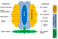
 Cerebellum Einteilung.png 914 × 637; 52 KB
-
 CerebellumDiv zh.png 914 × 637; 59 KB
-
 CerebellumDiv-es.png 1,000 × 697; 157 KB
-
 CerebellumDiv.png 914 × 637; 67 KB
-
 CerebellumDivRO.png 914 × 637; 52 KB
-
 Diseases of the nervous system (1908) (14592660687).jpg 2,096 × 1,664; 1.29 MB
-
 Diseases of the nervous system (1910) (14586285529).jpg 2,104 × 1,664; 571 KB
-
 Gray702 Cerebellar vermis.png 600 × 319; 308 KB
-
 Gray702.png 600 × 319; 73 KB
-
 Gray703 Cerebellar vermis.png 600 × 350; 226 KB
-
 Gray703.png 600 × 350; 59 KB
-
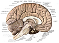
 Human cerebellum anterior view description.JPG 344 × 200; 16 KB
-
 Human cerebellum anterior view.JPG 344 × 200; 14 KB
-
 Human cerebellum posterior view description.JPG 345 × 211; 18 KB
-
 Human cerebellum posterior view.JPG 345 × 211; 16 KB
-
 Kleinhirn-es.png 1,600 × 1,099; 252 KB
-
 Kleinhirn.png 1,577 × 1,083; 124 KB
-
 Lawrence 1960 18.1.png 2,268 × 1,692; 1.02 MB
-
 Lawrence 1960 2.43.png 1,996 × 2,588; 1 MB
-
 Lawrence 1960 2.44.png 1,976 × 2,412; 863 KB
-
 Lawrence 1960 2.45.png 2,212 × 1,776; 932 KB
-
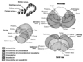
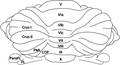 Lobules in the vermis.png 945 × 514; 183 KB
-
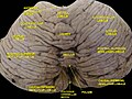
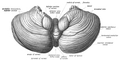
 PSM V26 D760 Upper surface of the cerebellum.jpg 1,247 × 915; 295 KB
-
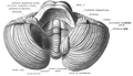
 PSM V26 D761 Inferior surface of the cerebellum.jpg 1,273 × 872; 310 KB
-
 Slide2AST-ar.jpg 960 × 720; 174 KB
-
 Slide2AST.JPG 960 × 720; 81 KB
-
 Slide2BRA-ar.jpg 960 × 720; 200 KB
-
 Slide2BRA.JPG 960 × 720; 103 KB
-
 Slide3AST.JPG 960 × 720; 92 KB
-
 Sobo 1909 624 ar.png 3,060 × 2,247; 5.38 MB
-
 Sobo 1909 624.png 3,060 × 2,247; 19.71 MB
-
 Sobo 1909 648.png 1,063 × 1,048; 3.19 MB
-
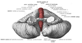
 Sobo 1909 653 Cerebellar vermis.png 1,004 × 512; 356 KB
-
 Sobo 1909 653.png 1,004 × 512; 1.47 MB
-
 Sobo 1909 654 Cerebellar vermis.png 1,007 × 507; 303 KB
-
 Sobo 1909 654.png 1,007 × 507; 1.46 MB
-
 Sobo 1909 655 Cerebellar vermis.png 1,022 × 539; 311 KB
-
 Sobo 1909 655.png 1,022 × 539; 1.58 MB
-
 Sobo 1909 656.png 867 × 498; 1.24 MB
-
Vermis and paravermis by Sanjoy Sanyal.webm 1 min 5 s, 718 × 404; 6.7 MB
-
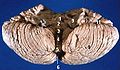
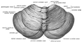
 Vermis.jpg 988 × 918; 65 KB


_(14592660687).jpg)
_(14586285529).jpg)


