Category:Histology of the human pineal gland
Media in category "Histology of the human pineal gland"
The following 55 files are in this category, out of 55 total.
-
 Blood Vessels in Septae of Older Human Pineal Gland by Phase Contrast (46684388894).jpg 3,264 × 1,840; 1.57 MB
-
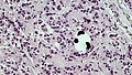
 Brain Sand in Older Human Pineal Gland (33527661718).jpg 3,264 × 1,840; 2.13 MB
-
 Brain Sand in Older Human Pineal Gland (40440641473).jpg 3,264 × 1,840; 5.03 MB
-
 Brain Sand in Older Human Pineal Gland (46491416155).jpg 3,264 × 1,840; 6.74 MB
-
 Brain Sand in Older Human Pineal Gland (47352928552).jpg 3,264 × 1,840; 1.91 MB
-
 Brain Sand in Older Human Pineal Gland (47353308772).jpg 3,264 × 1,840; 1.47 MB
-
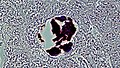 Brain Sand in Older Human Pineal Gland by Phase Contrast (46683386794).jpg 3,264 × 1,840; 2.48 MB
-
 Brain Sand in Older Human Pinealocyte by Phase Contrast (46491414525).jpg 3,264 × 1,840; 3.02 MB
-
 Brain Sand in Older Pineal Gland by Phase Contrast (46684389344).jpg 3,264 × 1,840; 1.91 MB
-
 Capillary Supply in Young Human Pineal Gland (40406904583).jpg 3,264 × 1,840; 2.21 MB
-

-
 Connective Tissue Wrapped Lobes in Older Human Pineal Gland (47352927622).jpg 3,264 × 1,840; 2.52 MB
-
 Connective Tissue Wrapped Lobes in Older PIneal Gland by Phase Contrast (46491415755).jpg 3,264 × 1,840; 3.02 MB
-
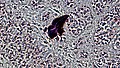
 Deposits of Sand in Older Human Pineal Gland by Phase Contrast (46683387694).jpg 3,264 × 1,840; 8.95 MB
-
 Human Pineal Gland Young (40406900493).jpg 3,264 × 1,840; 8.2 MB
-
 Human Pineal Gland Young (47319577772).jpg 3,264 × 1,840; 9.15 MB
-
 Human Pineal Gland Young (47372420061).jpg 3,264 × 1,840; 8.42 MB
-
 Human Pineal Gland Young by Phase Contrast (33496196008).jpg 3,264 × 1,840; 2.31 MB
-
 Human Pineal Gland Young by Phase Contrast (46457456425).jpg 3,264 × 1,840; 1.57 MB
-
 Human Pineal Gland Young by Phase Contrast (46457456745).jpg 3,264 × 1,840; 2.68 MB
-
 Human Pineal Gland Young by Phase Contrast (46457461585).jpg 3,264 × 1,840; 6.47 MB
-
 Large Brain Sand Deposit in Older Human Pineal Gland (40442211613).jpg 3,264 × 1,840; 1.24 MB
-
 Large Deposit of Brain Sand in Older Human Pineal by Phase Contrast (47354891072).jpg 3,264 × 1,840; 5.31 MB
-
 Older Human Pituitary Gland by Phase Contrast (40442211443).jpg 3,264 × 1,840; 1.55 MB
-
 Pia Mater in Older Human Pineal Gland by Phase Contrast (40442210013).jpg 3,264 × 1,840; 2.16 MB
-
 Pineal gland - high mag.jpg 2,848 × 4,272; 6.35 MB
-
 Pineal gland - intermed mag.jpg 2,848 × 4,272; 6.91 MB
-
 Pineal gland - low mag.jpg 2,848 × 4,272; 7.99 MB
-
 Pineal gland - very high mag.jpg 2,848 × 4,272; 5.36 MB
-
 Pineal.jpg 4,080 × 3,072; 3.13 MB
-
 Pinealocyte Rosettes in Older Pineal Gland (40440642443).jpg 3,264 × 1,840; 2.1 MB
-
 Pinealocytes and Associated Astrocytes in Older Pineal Gland (33530193708).jpg 3,264 × 1,840; 1.61 MB
-
 Pinealocytes and Astrocytes in Young Human Pineal Gland (40406903583).jpg 3,264 × 1,840; 7.26 MB
-

-
 Rosette of Pinealocytes in Young Human Pineal Gland (40406903783).jpg 3,264 × 1,840; 8.12 MB
-
 Rosettes of Pinealocytes in Older Human Pineal Gland (47352928292).jpg 3,264 × 1,840; 2.54 MB
-
 Rosettes of Pinealocytes in Older Human Pineal Gland by Phase Contrast (40441156793).jpg 3,264 × 1,840; 3.04 MB
-
 Rosettes of Pinealocytes in Older Human Pineal Gland by Phase Contrast (47354890892).jpg 3,264 × 1,840; 1.58 MB
-
 Rosettes of Pinealocytes in Young Human Pineal Gland by Phase Contrast (46457461285).jpg 3,264 × 1,840; 6.11 MB
-
 Septate Spaces Between Lobes in Older Human Pineal Gland by Phase Contrast (40441155733).jpg 3,264 × 1,840; 3.06 MB
-
 Tightly Adherent Pia Mater in Older Human Pineal Gland (40440641833).jpg 3,264 × 1,840; 1.87 MB
-
 Tightly Adherent Pia Mater in Older Human Pineal Gland (47352927992).jpg 3,264 × 1,840; 1.99 MB
-
 Well Defined Lobes in Older Human Pineal Gland (33527661368).jpg 3,264 × 1,840; 2.6 MB
-
 Well Defined Lobes in Older Human Pineal Gland by Phase Contrast (40442209533).jpg 3,264 × 1,840; 1.43 MB
-
 Young Pineal Gland Hematoxylin and Eosin Staining (33496198538).jpg 3,264 × 1,840; 1.38 MB
-
 Young Pineal Gland Hematoxylin and Eosin Staining (33496198688).jpg 3,264 × 1,840; 1.36 MB
-
 Young Pineal Gland Hematoxylin and Eosin Staining (33496198818).jpg 3,264 × 1,840; 1.38 MB
-
 Young Pineal Gland Hematoxylin and Eosin Staining (33496198968).jpg 3,264 × 1,840; 1.41 MB
-
 Young Pineal Gland Hematoxylin and Eosin Staining (33496199108).jpg 3,264 × 1,840; 1.75 MB
-
 Young Pineal Gland Hematoxylin and Eosin Staining (46457459845).jpg 3,264 × 1,840; 2.15 MB
-
 Young Pineal Gland Hematoxylin and Eosin Staining (46457460095).jpg 3,264 × 1,840; 1.77 MB
-
 Young Pineal Gland Hematoxylin and Eosin Staining (46457460375).jpg 3,264 × 1,840; 1.71 MB
-
 Young Pineal Gland Hematoxylin and Eosin Staining (46457460555).jpg 3,264 × 1,840; 1.81 MB
-
 Young Pineal Gland Hematoxylin and Eosin Staining (46457460735).jpg 3,264 × 1,840; 1.77 MB
-
 Young Pineal Gland Hematoxylin and Eosin Staining (46457461065).jpg 3,264 × 1,840; 1.59 MB
.jpg)
.jpg)
.jpg)
.jpg)
.jpg)
.jpg)
.jpg)
.jpg)
.jpg)
.jpg)
.jpg)
.jpg)
.jpg)
.jpg)
.jpg)
.jpg)
.jpg)
.jpg)
.jpg)
.jpg)
.jpg)
.jpg)
.jpg)
.jpg)
.jpg)


.jpg)
.jpg)
.jpg)
.jpg)
.jpg)
.jpg)
.jpg)
.jpg)
.jpg)
.jpg)
.jpg)
.jpg)
.jpg)
.jpg)
.jpg)
.jpg)
.jpg)
.jpg)
.jpg)
.jpg)
.jpg)
.jpg)
.jpg)
.jpg)
.jpg)