Category:Human neuroanatomy photographs
Media in category "Human neuroanatomy photographs"
The following 84 files are in this category, out of 84 total.
-
 Human base of brain blood supply description.JPG 501 × 540; 42 KB
-
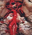
 Human base of brain blood supply.JPG 501 × 540; 38 KB
-
 Human brain anterior-inferior view description.JPG 330 × 475; 31 KB
-
 Human brain anterior-inferior view.JPG 330 × 475; 28 KB
-
 Human brain arachnoid description.JPG 699 × 480; 41 KB
-
 Human brain arachnoid.JPG 699 × 480; 39 KB
-

-
 Human brain central arteries anterior midsagittal view.JPG 701 × 482; 56 KB
-
 Human brain dura mater (reflections) description.JPG 700 × 486; 49 KB
-
 Human brain dura mater (reflections).JPG 700 × 486; 44 KB
-
 Human brain dura mater description.JPG 555 × 484; 44 KB
-
 Human brain dura mater.JPG 555 × 484; 41 KB
-

-
 Human brain frontal (coronal) section description 2.JPG 702 × 487; 43 KB
-
 Human brain frontal (coronal) section description.JPG 702 × 487; 43 KB
-
 Human brain frontal (coronal) section description2.JPG 702 × 487; 42 KB
-
 Human brain frontal (coronal) section.JPG 702 × 487; 41 KB
-
 Human brain inferior view description 2.JPG 373 × 467; 36 KB
-
 Human brain inferior view description.JPG 373 × 466; 37 KB
-
 Human brain inferior view.JPG 373 × 467; 34 KB
-

-
 Human brain inferior-medial view description 2.JPG 702 × 491; 68 KB
-
 Human brain inferior-medial view description 3.JPG 702 × 491; 63 KB
-
 Human brain inferior-medial view description.JPG 702 × 491; 63 KB
-
 Human brain inferior-medial view with marked Precuneus.JPG 702 × 491; 211 KB
-
 Human brain inferior-medial view.JPG 702 × 491; 52 KB
-
 Human brain lateral view cut-line brainstem.JPG 701 × 487; 48 KB
-
 Human brain lateral view description 2.JPG 701 × 487; 50 KB
-
 Human brain lateral view description.JPG 701 × 487; 49 KB
-
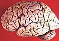
 Human brain lateral view.JPG 701 × 487; 43 KB
-
 Human brain left dissected midsagittal view description 2.JPG 701 × 488; 50 KB
-
 Human brain left dissected midsagittal view description.JPG 701 × 488; 50 KB
-
 Human brain left dissected midsagittal view.JPG 701 × 488; 46 KB
-

-
 Human brain left midsagitttal view closeup description 2.JPG 701 × 490; 61 KB
-
 Human brain left midsagitttal view closeup description 3.JPG 701 × 490; 58 KB
-
 Human brain left midsagitttal view closeup description.JPG 701 × 490; 58 KB
-
 Human brain left midsagitttal view closeup.JPG 701 × 490; 53 KB
-
 Human brain midsagittal cut color.png 1,294 × 861; 1.17 MB
-
 Human brain midsagittal cut color2.png 958 × 720; 1.14 MB
-
 Human brain midsagittal cut description.JPG 701 × 486; 50 KB
-
 Human brain midsagittal cut.JPG 701 × 486; 45 KB
-
 Human brain midsagittal view description.JPG 423 × 374; 30 KB
-
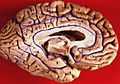
 Human brain midsagittal view.JPG 423 × 374; 27 KB
-
 Human brain right dissected lateral view description.JPG 653 × 413; 40 KB
-
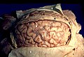
 Human brain right dissected lateral view.JPG 653 × 413; 37 KB
-

-
 Human brain right Great Cerebral Vein midsagittal view.JPG 691 × 439; 50 KB
-
 Human brain superior-lateral view description.JPG 489 × 332; 32 KB
-

-
 Human brain superior-lateral view.JPG 490 × 334; 30 KB
-

-
 Human brain view on transverse temporal and insular gyri.JPG 470 × 326; 22 KB
-
 Human brainstem anterior view 2 description.JPG 346 × 487; 35 KB
-
 Human brainstem anterior view 2.JPG 346 × 487; 30 KB
-
 Human brainstem anterior view blood supply description.JPG 335 × 466; 30 KB
-
 Human brainstem anterior view blood supply.JPG 335 × 466; 30 KB
-
 Human brainstem anterior view description 2.JPG 347 × 485; 31 KB
-
 Human brainstem anterior view description.JPG 347 × 485; 31 KB
-
 Human brainstem anterior view.JPG 347 × 485; 27 KB
-
 Human brainstem blood supply description 2.JPG 330 × 467; 33 KB
-
 Human brainstem blood supply description.JPG 331 × 468; 36 KB
-
 Human brainstem blood supply.JPG 331 × 468; 32 KB
-
 Human brainstem-thalamus posterior view description.JPG 340 × 485; 23 KB
-
 Human brainstem-thalamus posterior view.JPG 342 × 487; 20 KB
-
 Human brainstem-thalamus posterior-inferior view description.JPG 405 × 487; 26 KB
-
 Human brainstem-thalamus posterior-inferior view.JPG 405 × 487; 24 KB
-
 Human caudal brainstem posterior view description.JPG 344 × 475; 20 KB
-
 Human caudal brainstem posterior view.JPG 344 × 475; 17 KB
-
 Human caudal spinal cord anterior view description.jpg 229 × 468; 26 KB
-
 Human caudal spinal cord anterior view.jpg 229 × 468; 24 KB
-
 Human cerebellum anterior view description.JPG 344 × 200; 16 KB
-
 Human cerebellum anterior view.JPG 344 × 200; 14 KB
-
 Human cerebellum posterior view description.JPG 345 × 211; 18 KB
-
 Human cerebellum posterior view.JPG 345 × 211; 16 KB
-

-
 Human cerebrum lateral view, a part of temporal lobe resected.JPG 699 × 491; 50 KB
-
 Human spinal cord anterior view description.jpg 661 × 461; 29 KB
-
 Human spinal cord anterior view.jpg 661 × 461; 28 KB
-
 Human spinal cord posterior and anterior view description.jpg 699 × 486; 38 KB
-
 Human spinal cord posterior and anterior view.jpg 699 × 486; 34 KB
-
 Location of Midbrain in inferior view.png 373 × 467; 307 KB
-
 Nervous and mental diseases (1908) (14755550006).jpg 1,912 × 1,348; 686 KB
-
 Skull inner surface.jpg 2,409 × 3,134; 5.27 MB


_description.JPG)
.JPG)


_section_description_2-emphasizing-corpus-callosum.png)
_section_description_2.JPG)
_section_description.JPG)
_section_description2.JPG)
_section.JPG)


_(14755550006).jpg)
