Category:Microscopic images of animal bones
Media in category "Microscopic images of animal bones"
The following 23 files are in this category, out of 23 total.
-

-

-
 Aenigmastropheus histology.png 1,328 × 1,598; 4.91 MB
-
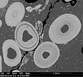
 Bertazzo S SEM deproteined bone - cranium rat - x10k.jpg 1,280 × 960; 686 KB
-
Blood-flow-controls-bone-vascular-function-and-osteogenesis-ncomms13601-s2.ogv 2.9 s, 1,921 × 1,325; 8.67 MB
-
 Bone (1).jpg 3,164 × 2,151; 1.64 MB
-
 Bone Cells (10835380615).jpg 355 × 273; 48 KB
-
 Bone histology of Shuvuuia and Confuciusornis.png 1,240 × 880; 1.5 MB
-
 Bone histomorphometry.jpg 2,048 × 1,536; 4.33 MB
-
 Bone marrow cow horizontal.jpg 600 × 432; 51 KB
-
 Bone marrow cow.jpg 600 × 421; 41 KB
-
 Collage of fish bones.jpg 2,202 × 1,791; 1.2 MB
-
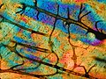 Dünnschliff Rinderknochen Metatarsus pol.jpg 1,600 × 1,200; 701 KB
-
 Fossil-AvimaiaSchweitzerae-Histology-MedullaryBone.png 1,350 × 1,039; 1.88 MB
-
 Hemidactylus (10.3897-zse.94.22289) Figure 6.jpg 1,775 × 2,539; 2.88 MB
-
 Limb bone osteohistology of Brasilitherium riograndensis.png 4,296 × 5,503; 29.32 MB
-
 Marrow Adipose Tissue (typical quantity young mouse) .jpg 10,200 × 13,200; 4.93 MB
-
 Marrow Adipose Tissue (typical quantity young mouse) cropped.jpg 587 × 590; 503 KB
-
 NSMT-PL 570 7.18 mm frontal.jpg 1,242 × 846; 176 KB
-
 Ooidal ironstone.jpg 1,024 × 943; 131 KB
-
 Probrachylophosaurus histology.PNG 1,602 × 2,179; 6.65 MB
-
 Proximal tibia Masson Goldner Trikrom rabbit 600x growth zone.jpg 800 × 600; 793 KB
-
 Whirling disease pathology.jpg 1,099 × 736; 485 KB
_(14596927200).jpg)


.jpg)
.jpg)


_Figure_6.jpg)

_.jpg)
_cropped.jpg)


