Category:Microscopy images from Wiki Science Competition 2023 in Ukraine
Media in category "Microscopy images from Wiki Science Competition 2023 in Ukraine"
The following 43 files are in this category, out of 43 total.
-
 Adenocarcinoma of the prostate.jpg 4,096 × 3,008; 2.27 MB
-
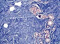
 Amyloid deposition in prostate tumor tissue.jpg 4,096 × 3,008; 2.15 MB
-
 Anaphase meiosis I.png 1,392 × 1,024; 1.28 MB
-
 Composite for hydrogen storage.png 2,048 × 1,144; 3.5 MB
-
 Cross-section through the spinal cord.jpg 4,205 × 2,977; 5.6 MB
-
 Differentiation of Human-Induced Pluripotent Stem Cells to GABAergic Neurons.tif 1,296 × 966; 3.58 MB
-
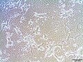
 Du145 cell line.jpg 2,048 × 1,536; 360 KB
-
 Fibrin clot formed in the presence of F(ab) fragments of monoclonal antibody III-1D.jpg 1,696 × 2,090; 723 KB
-
 Immunohistochemistry technologies with AI.png 730 × 346; 419 KB
-
 Invasive ductal carcinoma.jpg 4,096 × 3,008; 2.38 MB
-
 Invasive lobular carcinoma.jpg 4,096 × 3,008; 2.58 MB
-
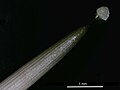
 Iris sp. root cross-sectional preparation in the microscope.jpg 1,080 × 1,080; 141 KB
-
 L929 cell line.jpg 2,048 × 1,536; 582 KB
-
 Layers of stomach.jpg 3,273 × 2,574; 5.8 MB
-
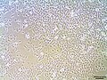
 LnCup cell line.jpg 2,048 × 1,536; 496 KB
-
 MCF7 cell line.jpg 2,048 × 1,536; 532 KB
-
 MDA BM 231 cell line.jpg 2,048 × 1,536; 450 KB
-
 Metaphase meiosis II.png 1,392 × 1,024; 1.15 MB
-
 Metephase meiosis I.jpg 1,050 × 800; 259 KB
-
 Microscopic image of a pumpkin stem cell.jpg 5,715 × 5,116; 24.6 MB
-
 Nanoart Bavovna.jpg 2,080 × 1,560; 1.31 MB
-
 Nanoart Exoplanets.jpg 2,048 × 1,755; 1,007 KB
-
 Nanoart Hope.jpg 2,080 × 1,560; 1.26 MB
-
 Nanoart Leopard.jpg 2,080 × 1,560; 1.92 MB
-
 Nanoart Lightning of Victory.jpg 4,096 × 3,072; 3.27 MB
-
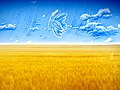
 Nanoart One Love Ukraine.jpg 1,560 × 2,080; 2.38 MB
-
 Neuron in the microchannel of hydrogel implant.png 5,240 × 1,034; 9.97 MB
-
 Neutral red uptake assay. Rats embrionic fibroblasts.jpg 2,048 × 1,536; 381 KB
-
 PC3 cell line.jpg 2,048 × 1,536; 443 KB
-
 Pikseli ekrana telefona.jpg 2,999 × 1,982; 789 KB
-
 PK15 cell line.jpg 2,048 × 1,536; 366 KB
-
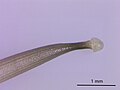
 Pteridium aquilinum rhizome.jpg 4,280 × 3,400; 8.62 MB
-
 Scanning electron micrograph of mycosynthesized ZnO nanoflowers.jpg 1,024 × 1,144; 62 KB
-
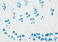 Small intestine Alcian blue 400x.jpg 4,096 × 3,008; 1.4 MB
-
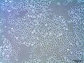
 SPEV cell line.jpg 2,048 × 1,536; 513 KB
-
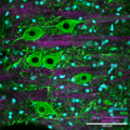 Spinal cord gray matter immunofluorescence staining, confocal imaging.png 1,600 × 1,600; 5.88 MB
-
 Spongy bone in polarized light.jpg 2,480 × 1,419; 3.65 MB
-
 T47D cell line.jpg 2,048 × 1,536; 534 KB
-
 Telophase meiosis I.png 1,392 × 1,024; 1.28 MB
-
 Toxoplasma parasites (tachyzoites) in a fibroblast host cell.png 1,254 × 1,254; 1.08 MB
-
-
 Мегакаріоцит в червоному кістковому мозку.tif 3,840 × 2,160; 5.08 MB
-


_fragments_of_monoclonal_antibody_III-1D.jpg)


_in_a_fibroblast_host_cell.png)
.jpg)
.jpg)