Category:SVG human brain (sagittal section)
Media in category "SVG human brain (sagittal section)"
The following 60 files are in this category, out of 60 total.
-
 202102 Mid-sagittal plane of the brain.svg 512 × 512; 2.72 MB
-
 Arbor Vitae in the Human Cerebellum.svg 512 × 399; 91 KB
-
 Blank Diagram of the Human Cerebellum.svg 512 × 399; 92 KB
-
 Brain anatomy.svg 512 × 494; 1.61 MB
-
 Brain bulbar region Ar.svg 295 × 299; 217 KB
-
 Brain bulbar region as.svg 295 × 299; 196 KB
-
 Brain bulbar region IT.svg 295 × 299; 195 KB
-
 Brain bulbar region ja.svg 295 × 299; 111 KB
-
 Brain bulbar region ml.svg 295 × 299; 195 KB
-
 Brain bulbar region ta.svg 295 × 299; 196 KB
-
 Brain bulbar region-es.svg 295 × 299; 134 KB
-
 Brain bulbar region-gu.svg 295 × 299; 195 KB
-
 Brain bulbar region-mr.svg 295 × 299; 196 KB
-
 Brain bulbar region-te.svg 295 × 299; 194 KB
-
 Brain bulbar region-ur.svg 295 × 299; 194 KB
-
 Brain bulbar region.PNG 295 × 299; 51 KB
-
 Brain bulbar region.svg 295 × 299; 194 KB
-
 Brain bulbar region.svg-kn.svg 295 × 299; 208 KB
-
 Brain human sagittal section.svg 295 × 326; 145 KB
-
 Brain logo.svg 612 × 792; 38 KB
-
 Brain sagittal section stem highlighted.svg 295 × 326; 152 KB
-
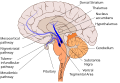
 Brain stem sagittal section.svg 566 × 639; 355 KB
-
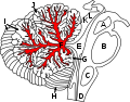
 Cerebral vascular territories midline.svg 512 × 481; 74 KB
-
 CorpusCallosum.svg 1,025 × 598; 25 KB
-
 Dopaminergic pathways.svg 446 × 307; 185 KB
-
 Encephalon human sagittal section multilingual.svg 401 × 374; 158 KB
-
 Fourth Ventricle in the Human Cerebellum.svg 512 × 399; 129 KB
-
 Gehirn, medial - beschriftet lat-rus.svg 632 × 477; 499 KB
-
 Gehirn, medial - beschriftet lat.svg 632 × 477; 307 KB
-
 Gehirn, medial - Lobi ar.svg 724 × 482; 96 KB
-
 Gehirn, medial - Lobi deu.svg 739 × 482; 121 KB
-
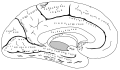
 Gehirn, medial - Lobi en.svg 724 × 482; 120 KB
-
 Gehirn, medial - Lobi es.svg 724 × 482; 55 KB
-
 Gehirn, medial - Lobi ja.svg 724 × 482; 53 KB
-
 Gray727 latin.svg 1,025 × 598; 23 KB
-
 Gray727.svg 1,025 × 598; 18 KB
-
 Head anatomy lateral view.svg 1,344 × 1,360; 57 KB
-
 Human brain - Sagittal section.svg 380 × 400; 170 KB
-
 Inferior colliculus of the human midbrain.svg 512 × 399; 92 KB
-
 Medulla Oblongata and Cerebellum.svg 512 × 399; 91 KB
-
 Mesocortical pathway.svg 446 × 307; 167 KB
-
 Mesolimbic pathway.svg 446 × 307; 167 KB
-
 Midbrain of the Human Brainstem.svg 512 × 399; 91 KB
-
 Nigrostriatal pathway.svg 446 × 307; 167 KB
-
 Pituitary gland representation cs.svg 261 × 236; 6 KB
-
 Pituitary gland representation es.svg 261 × 236; 8 KB
-
 Pituitary gland representation fr.svg 261 × 236; 5 KB
-
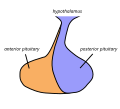 Pituitary gland representation.svg 261 × 236; 6 KB
-
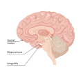
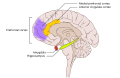 PTSD brain.svg 530 × 342; 149 KB
-
 Serotonergic neurons Ar.svg 295 × 299; 229 KB
-
 Serotonergic neurons.svg 295 × 299; 205 KB
-
 Skull and brain sagittal uk.svg 510 × 402; 470 KB
-
 Skull and brain sagittal.svg 218 × 219; 240 KB
-
 Skull and brainstem inner ear.svg 574 × 612; 702 KB
-
 Skull and sagittal brain.svg 411 × 537; 157 KB
-
 Superior colliculus of the human midbrain.svg 512 × 399; 92 KB
-
 Transsphenoidaler Zugang.svg 617 × 524; 330 KB
-
 Tuberoinfundibular pathway.svg 446 × 307; 169 KB
-
 Ventral tegmental area.svg 446 × 307; 174 KB
-
 ヒトの脳 模式図ii.svg 1,052 × 744; 41 KB


