Category:SVG mitosis
Subcategories
This category has only the following subcategory.
Media in category "SVG mitosis"
The following 107 files are in this category, out of 107 total.
-
 Activities of key regulators during entry into mitosis.svg 550 × 498; 61 KB
-
 Anaphase eukaryotic mitosis.svg 994 × 699; 146 KB
-
 Anaphase.svg 2,680 × 1,400; 174 KB
-
 Animal cell cycle (editors version).svg 7,207 × 7,003; 752 KB
-
 Animal cell cycle - eu.svg 7,207 × 7,003; 1.17 MB
-
 Animal cell cycle ar.svg 7,207 × 7,003; 1.37 MB
-
 Animal cell cycle-ar.svg 7,207 × 7,003; 1.46 MB
-
 Animal cell cycle-en.svg 7,207 × 7,003; 1.45 MB
-
 Animal cell cycle-es.svg 7,207 × 7,003; 1.48 MB
-
 Animal cell cycle-fr.svg 7,790 × 6,883; 1.69 MB
-
 Animal cell cycle-ku.svg 7,688 × 7,469; 731 KB
-
 Animal cell cycle-pt-br.svg 512 × 497; 355 KB
-
 Animal cell cycle-tr.svg 512 × 497; 360 KB
-
 Animal cell cycle-zh-hant.svg 512 × 497; 1.25 MB
-
 APC (ru).svg 473 × 160; 25 KB
-
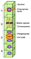
 Chromosomes during mitosis.svg 285 × 380; 145 KB
-
 Citocinesis de mitosis eucariota.svg 994 × 699; 216 KB
-
 Cytokinesis eukaryotic mitosis.svg 994 × 699; 120 KB
-
 Diagram of mitosis.svg 477 × 337; 252 KB
-
 Diagram showing mitosis CRUK 411.svg 375 × 225; 48 KB
-
 Diploidisolu.svg 180 × 303; 3 KB
-
 Evolution of mitosis in protozoa.svg 1,081 × 1,536; 487 KB
-
 Filmstreifen Mitose.svg 512 × 2,501; 38 KB
-
 First gap cell lifecycle.svg 994 × 699; 112 KB
-
 Fungus cell cycle-en.svg 2,310 × 1,491; 2.74 MB
-
 Fus-acromatic.svg 580 × 410; 25 KB
-
 Fus-astral-anastral.svg 3,100 × 2,800; 605 KB
-
 Hauptereignisse der Mitose.svg 1,023 × 372; 11 KB
-
 Interphase and Mitosis.svg 1,000 × 350; 80 KB
-
 Les 4 phases principales de la mitose.svg 785 × 740; 23 KB
-
 Major events in mitosis be.svg 1,023 × 372; 15 KB
-
 Major events in mitosis ca.svg 1,023 × 372; 36 KB
-
 Major events in mitosis el.svg 1,023 × 372; 11 KB
-
 Major events in mitosis he.svg 1,023 × 372; 11 KB
-
 Major events in mitosis ml.svg 1,127 × 488; 17 KB
-
 Major events in mitosis NL.svg 1,023 × 372; 10 KB
-
 Major events in mitosis ru.svg 1,023 × 372; 15 KB
-
 Major events in mitosis sk.svg 1,023 × 372; 10 KB
-
 Major events in mitosis sl.svg 1,023 × 372; 10 KB
-
 Major events in mitosis-gl.svg 1,023 × 372; 10 KB
-
 Major events in mitosis.svg 1,023 × 372; 7 KB
-
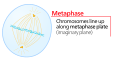
 Metaphase eukaryotic mitosis.svg 994 × 699; 125 KB
-
 Metaphase.svg 2,680 × 1,400; 137 KB
-
 Mitoosi1.svg 722 × 440; 5 KB
-
 Mitoosi2.svg 878 × 489; 8 KB
-
 Mitosis cells sequence.svg 774 × 115; 459 KB
-
 Mitosis classification closed extranuclear pleuromitoses.svg 187 × 109; 78 KB
-
 Mitosis classification closed intranuclear pleuromitoses.svg 187 × 109; 8 KB
-
 Mitosis classification closed orthomitoses.svg 187 × 109; 13 KB
-
 Mitosis classification open orthomitoses.svg 187 × 109; 20 KB
-
 Mitosis classification semiopen orthomitoses.svg 187 × 109; 39 KB
-
 Mitosis classification semiopen pleuromitoses.svg 187 × 109; 10 KB
-
 Mitosis diagram.jpg 1,422 × 1,050; 269 KB
-
 Mitosis schematic diagram ku.svg 833 × 723; 208 KB
-
 Mitosis schematic diagram-en.svg 833 × 723; 192 KB
-
 Mitosis Stages - Numerical version.svg 2,361 × 409; 787 KB
-
 Mitosis Stages - Persian.svg 2,361 × 409; 1.07 MB
-
 Mitosis Stages ku.svg 2,361 × 409; 1.16 MB
-
 Mitosis Stages.svg 2,361 × 409; 1.13 MB
-
 Mitosis.svg 744 × 1,052; 21 KB
-
 Mitotic Anaphase.svg 120 × 78; 77 KB
-
 Mitotic crossing-over.svg 512 × 456; 99 KB
-
 Mitotic Cytokinesis.svg 150 × 92; 148 KB
-
 Mitotic Metaphase.svg 97 × 81; 78 KB
-
 Mitotic Prometaphase.svg 79 × 72; 75 KB
-
 Mitotic Prophase.svg 76 × 80; 89 KB
-
 Mitotic spindle in animal cell.svg 645 × 573; 192 KB
-
 Mitotic spindle in budding yeast.svg 701 × 548; 172 KB
-
 Mitotic spindle in plant cell.svg 645 × 573; 192 KB
-
 Mitotic Telophase.svg 129 × 80; 131 KB
-
 Mitoza.svg 1,169 × 826; 197 KB
-

-
 Overview of mitotic control mechanisms (ru).svg 651 × 384; 51 KB
-
 Phase of mitosis (ru).svg 1,449 × 3,407; 256 KB
-
 Plant cell cycle.svg 1,440 × 1,240; 3.51 MB
-
 Plant cell cytokinesis.svg 580 × 400; 51 KB
-
 Plant cell division in a cell with a large vacuole.svg 512 × 1,037; 53 KB
-
 Plant cell early anaphase.svg 580 × 380; 91 KB
-
 Plant cell first gap.svg 580 × 400; 20 KB
-
 Plant cell late anaphase.svg 580 × 380; 105 KB
-
 Plant cell metaphase.svg 580 × 380; 86 KB
-
 Plant cell prometaphase.svg 580 × 380; 68 KB
-
 Plant cell prophase.svg 580 × 380; 74 KB
-
 Plant cell S phase and second gap.svg 580 × 385; 41 KB
-
 Plant cell telophase.svg 580 × 400; 152 KB
-
 Preprophaseband.png 412 × 786; 23 KB
-
 Prometaphase eukaryotic mitosis.svg 994 × 699; 165 KB
-
 Prometaphase.svg 2,680 × 1,400; 252 KB
-
 Prophase eukaryotic mitosis.svg 994 × 699; 198 KB
-
 Prophase.svg 2,680 × 1,400; 145 KB
-
 Schemazeichnung Mitose.svg 833 × 723; 183 KB
-
 Second gap cell lifecycle.svg 994 × 699; 130 KB
-
 Self-organization of a bipolar microtubule array.svg 498 × 1,264; 266 KB
-
 Spindle apparatus-es.svg 576 × 402; 16 KB
-
 Spindle apparatus.svg 576 × 402; 10 KB
-
 Spindle assembly models.svg 403 × 846; 51 KB
-
 Synthesis cell lifecycle.svg 994 × 699; 107 KB
-
 Telophase eukaryotic mitosis.svg 994 × 699; 166 KB
-
 Telophase.svg 2,880 × 1,800; 255 KB
-
 The Cell Cycle.svg 379 × 378; 32 KB
-
 The main types of mitosis in algae.svg 697 × 138; 60 KB
-
 The time course of mitosis and cytokinesis (ru).svg 548 × 629; 23 KB
-
 Three cell growth types-ru.svg 694 × 700; 241 KB
-
 Three cell growth types.svg 694 × 700; 284 KB
-
 Types of mitosis int.svg 700 × 600; 51 KB
-
 Événements importants en mitose.svg 1,023 × 372; 16 KB
-
 Митоза (анимација).gif 865 × 579; 3.01 MB


.svg)


.svg)
.svg)


.svg)


.gif)
.svg)