Category:Three-dimensional reconstructions with computed tomography images
Subcategories
This category has the following 4 subcategories, out of 4 total.
3
- 3D scans of fossils (32 F)
M
Media in category "Three-dimensional reconstructions with computed tomography images"
The following 139 files are in this category, out of 139 total.
-
 12-06-11-rechtsmedizin-berlin-07.jpg 2,021 × 2,014; 1.24 MB
-

-
 3D CT mandible fracture.jpg 653 × 628; 292 KB
-
 3D CT of bilateral mandible fracture.jpg 666 × 684; 349 KB
-
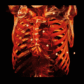
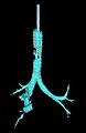
 3D CT of pulmonary circulation.jpg 1,082 × 1,155; 649 KB
-
 3D CT of thorax, annotated.jpg 1,544 × 1,131; 656 KB
-
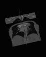
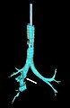
 3D CT of thorax.jpg 1,082 × 1,081; 555 KB
-
 3D CT of thorax.svg 1,544 × 1,131; 3.56 MB
-
 3D CT Reconstruction of Distal tibia fracture.gif 988 × 918; 9.32 MB
-
 3D model of human male and female pelvis.stl 5,120 × 2,880; 47.4 MB
-
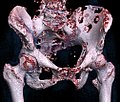
 3D rendered CT of bony pelvis 1.jpg 739 × 485; 122 KB
-
 3D rendered CT of bony pelvis 2.jpg 679 × 540; 141 KB
-
 3D rendered CT of bony pelvis 3.jpg 464 × 522; 90 KB
-
 3D rendered CT of hip bone metastases.jpg 1,003 × 850; 302 KB
-
 3D SSD.gif 256 × 256; 2.05 MB
-
 3D surface rendering CT 04 edited-1.jpg 480 × 497; 95 KB
-
-
 3DPX-002591 CE Abdominal CTA Nevit Dilmen.stl 5,120 × 2,880; 4.9 MB
-
 3DPX-002736 Large intestine Nevit Dilmen.stl 5,120 × 2,880; 20.91 MB
-
 3DPX-003169 Baby cranium NevitDilmen.stl 5,120 × 2,880; 31.51 MB
-
 3DPX-008679 Foot Skin Nevit Dilmen.stl 5,120 × 2,880; 22.31 MB
-
 AAA 40.8 mm.jpg 2,373 × 2,289; 1.19 MB
-
 AneurysmAorta.jpg 440 × 493; 68 KB
-
 AneurysmAortaWithArrows.jpg 440 × 493; 72 KB
-
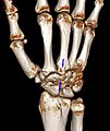
 Bilateral CT after hand transplant.jpg 666 × 479; 58 KB
-
 Bonereconstruction.jpg 260 × 259; 10 KB
-
 Bronchial stenosis CT.JPG 238 × 294; 6 KB
-
 Carpal-boss-3d.jpg 918 × 1,095; 179 KB
-
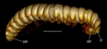
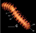
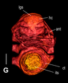
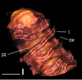
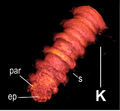
 Carychium hardiei (10.3897-zookeys.675.12453) Figure 5.jpg 1,512 × 1,749; 695 KB
-
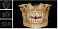 CBCT image 02.png 1,597 × 811; 1.32 MB
-
 Cbct skull.jpg 538 × 614; 52 KB
-
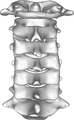
 Cervical spine Anterior.png 1,200 × 1,960; 1.21 MB
-
 ClutComp.jpg 522 × 522; 49 KB
-
 ClutCompos.jpg 517 × 520; 47 KB
-
 Coarctation of the aorta.tiff 984 × 307; 451 KB
-
 Computer tomography of a Norway Spruce cone 07.jpg 1,729 × 1,435; 770 KB
-
 Computer tomography of a pine cone 01.jpg 1,729 × 1,435; 622 KB
-
 Computer tomography of a pine cone 03.jpg 1,729 × 1,435; 737 KB
-
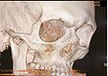
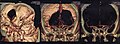
 Computer tomography Skull.jpg 1,128 × 805; 74 KB
-
 ContEnhcCompos.jpg 780 × 519; 56 KB
-
 Crane scann.JPG 1,133 × 798; 193 KB
-
 CT 3D reconstruction image of cervical spine Posterior view.png 1,200 × 1,954; 1.2 MB
-
 CtOrthor.gif 256 × 322; 926 KB
-
 CTSkullImage - cropped.jpg 768 × 600; 62 KB
-
 CTSkullImage.png 580 × 580; 336 KB
-
 CtVRvascRed.gif 256 × 256; 2.18 MB
-
 Disposable salt mill, computed tomogram (1).jpg 1,725 × 1,435; 156 KB
-
 Disposable salt mill, computed tomogram (2).jpg 1,725 × 1,435; 246 KB
-
 Disposable salt mill, computed tomogram (3).jpg 1,725 × 1,435; 387 KB
-
 Disposable salt mill, computed tomogram (4).jpg 1,725 × 1,435; 385 KB
-
 Disposable salt mill, computed tomogram (5).jpg 1,725 × 1,435; 268 KB
-
 Dual Energy CT images with gout tophi shown in green.jpg 750 × 805; 136 KB
-
 DVT Ansicht 1.png 886 × 591; 253 KB
-
 Glucose meter assembly.png 820 × 674; 178 KB
-
 Haidomyrmex scimitarus AMNH-BUFB80 02.jpg 1,286 × 808; 269 KB
-
 Haidomyrmex scimitarus AMNH-BUFB80 03.jpg 1,623 × 808; 290 KB
-
 Haidomyrmex scimitarus AMNH-BUFB80 04.jpg 727 × 808; 148 KB
-
 Haidomyrmex scimitarus AMNH-BUFB80 05.jpg 801 × 808; 253 KB
-
 Haidomyrmex scimitarus AMNH-BUFB80 06.jpg 1,174 × 808; 278 KB
-
 Horizontal Impaction.jpg 3,298 × 2,638; 2.43 MB
-
 Impacted mandibular canine.Gif 522 × 402; 4.35 MB
-
 Impacted wisdom tooth.gif 568 × 418; 225 KB
-
 Industrial Computed Tomography - Failures - Void.jpg 669 × 881; 176 KB
-
 Interspinous and intertuberous diameters.png 679 × 540; 589 KB
-
 Lumbar Vertebrae L3-L5 3D SR Nevit Dilmen.stl 5,120 × 2,880; 4.11 MB
-
 Massospondylus skull BP-1-5241 3D scan.stl 5,120 × 2,880; 2.37 MB
-
 Mediastinum.jpg 1,082 × 1,155; 641 KB
-
 Michelangelo.PNG 381 × 325; 116 KB
-
-
 Obstetric conjugate and pelvic outlet.png 464 × 522; 394 KB
-
 Odd couple.png 1,702 × 924; 2.65 MB
-
 OpacCompos.jpg 519 × 517; 52 KB
-
 Parry Romberg syndrome CT reconstruction, bone.jpg 1,054 × 1,003; 195 KB
-
 Parry Romberg syndrome CT reconstruction, soft tissues.jpg 1,093 × 936; 252 KB
-
 Pelvis CT 3d front 01.jpg 960 × 720; 52 KB
-
 Pelvis RG MR CT3d front 01.jpg 417 × 944; 44 KB
-
 Penetrating skull fracture.jpg 326 × 536; 23 KB
-
 PET physiologic muscle activity.jpg 1,024 × 1,024; 120 KB
-
 Pharmaceutical capsule, microtomography (1).jpg 1,761 × 1,435; 207 KB
-
 Pharmaceutical capsule, microtomography (2).jpg 1,761 × 1,435; 224 KB
-
 Pharmaceutical capsule, microtomography (3).jpg 1,761 × 1,435; 510 KB
-
 Pharmaceutical capsule, microtomography (4).jpg 1,761 × 1,435; 258 KB
-
 Pharmaceutical capsule, microtomography (5).jpg 1,761 × 1,435; 300 KB
-
 Pharmaceutical capsule, microtomography (6).jpg 1,761 × 1,435; 290 KB
-
 Pharmaceutical capsule, microtomography (7).jpg 1,761 × 1,435; 583 KB
-
 Plos One e105877 Figure 3 whole Maatidesmus paachtun.png 2,503 × 3,272; 9.81 MB
-
 Plos One e105877 Figure 3-A Maatidesmus paachtun.png 1,595 × 728; 740 KB
-
 Plos One e105877 Figure 3-B Maatidesmus paachtun.png 852 × 1,248; 803 KB
-
 Plos One e105877 Figure 3-C Maatidesmus paachtun.png 1,068 × 960; 486 KB
-
 Plos One e105877 Figure 3-D Maatidesmus paachtun.png 928 × 876; 442 KB
-
 Plos One e105877 Figure 3-E Maatidesmus paachtun.png 672 × 938; 352 KB
-
 Plos One e105877 Figure 3-F Maatidesmus paachtun.png 922 × 1,008; 508 KB
-
 Plos One e105877 Figure 3-G Maatidesmus paachtun.png 940 × 1,148; 577 KB
-
 Plos One e105877 Figure 3-H Maatidesmus paachtun.png 996 × 868; 569 KB
-
 Plos One e105877 Figure 3-I Maatidesmus paachtun.png 910 × 896; 516 KB
-
 Plos One e105877 Figure 3-K Maatidesmus paachtun.png 742 × 686; 241 KB
-
 Radiuskoepfchenfraktur-VR3.jpg 1,024 × 1,024; 433 KB
-
 RadiuskoepfchenfrakturVR2.png 1,024 × 1,024; 352 KB
-
 Rib fracture cystic fibrosis.jpg 1,404 × 1,875; 547 KB
-
 Rippenserienfraktur 3D CT.PNG 756 × 603; 630 KB
-
 Rotational micro-CT scan of Rhombophryne vaventy holotype.gif 784 × 732; 8.64 MB
-
 Severely impacted wisdom tooth CT scan - upper right.jpg 953 × 947; 134 KB
-
 ShadCompos.jpg 781 × 518; 83 KB
-
 Skull 3D SR File Nevit Dilmen.stl 5,120 × 2,880; 7.15 MB
-
 SPECT-CT of radioactive nanoparticles in mouse.png 2,059 × 2,031; 2.48 MB
-
 Ssd2surfs.jpg 508 × 565; 41 KB
-
 Swyer-James-Syndrom.jpg 551 × 551; 59 KB
-
 Thorax CT pcor Cut 3D Volume Rendering Haut.jpg 326 × 276; 13 KB
-
 Tomogram głowy u dorosłego człowieka.jpg 2,353 × 856; 3.48 MB
-
 Trabecular Bone Mouse Compared Spaceflight And Ground.jpg 314 × 159; 34 KB
-
 Tracheobronchial rupture 3D CT 2.jpg 239 × 369; 7 KB
-
 Tracheobronchial rupture 3D CT 3.JPG 239 × 369; 6 KB
-
 Tracheobronchial rupture 3D CT 3.jpg 239 × 369; 7 KB
-
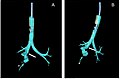 Tracheobronchial rupture 3D CT.jpg 600 × 397; 30 KB
-
 Ureterstent double J 3D legend.jpg 458 × 458; 37 KB
-
-

-
 Viewer medecine nucleaire keosys.JPG 1,440 × 900; 165 KB
-
 Violine3D Phillips.JPG 1,016 × 964; 112 KB
-
 Vocal folds cross section.jpg 549 × 586; 18 KB
-
-
 Volume rendered CT scan of abdominal and pelvic blood vessels (smaller).gif 644 × 862; 45.23 MB
-
 Volume rendered CT scan of abdominal and pelvic blood vessels.gif 822 × 1,100; 62.98 MB
-
 Volume rendering of abdominal muscles.jpg 771 × 943; 219 KB
-
 Volume rendering of hepatic veins, annotated.jpg 1,784 × 1,606; 666 KB
-
 Volume rendering of hepatic veins, annotated.svg 892 × 803; 1.41 MB
-
 Volume rendering of hepatic veins.jpg 892 × 803; 241 KB
-
 VrCrop.jpg 321 × 383; 20 KB
-
 Wall thickness analysis 2.png 152 × 160; 42 KB
-
 ΜCT Cupressus strobilus 01.jpg 942 × 1,232; 769 KB
-
 ΜCT Cupressus strobilus 02 - 3D section series.jpg 1,856 × 11,588; 7.32 MB
-
 ΜCT Cupressus strobilus 03 semi-transparent.jpg 1,040 × 1,232; 730 KB
-
 ΜCT Cupressus strobilus 04 mid section.jpg 1,856 × 1,435; 1.55 MB
-
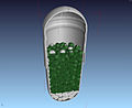
 ΜCT Kinder Schokobon 01.jpg 1,813 × 1,451; 947 KB
-
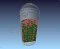
 ΜCT Kinder Schokobon 02.jpg 1,813 × 1,451; 970 KB
-
 ΜCT Kinder Schokobon 03.jpg 1,813 × 1,451; 1.29 MB
-
 ΜCT Kinder Schokobon 04.jpg 1,813 × 1,451; 1.85 MB
-
 ΜCT Kinder Schokobon 05.jpg 1,813 × 1,451; 942 KB
-
ΜCT of a cupressus strobilus, spiral 3D flight.ogv 20 s, 800 × 608; 10.08 MB


_Figure_5.jpg)


.jpg)
.jpg)
.jpg)
.jpg)
.jpg)


.jpg)
.jpg)
.jpg)
.jpg)
.jpg)
.jpg)
.jpg)


.gif)
.gif)


