Category:Valid SVG created with ChemSketch
Media in category "Valid SVG created with ChemSketch"
The following 150 files are in this category, out of 150 total.
-
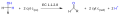
 (carboxyethyl)arginine beta-lactam-synthase.svg 561 × 188; 37 KB
-
 1,2-Thiazinane.svg 162 × 152; 3 KB
-
 1,3-Thiazinane.svg 162 × 153; 3 KB
-
 1,4-Dichlorobenzene 2.svg 248 × 166; 4 KB
-
 1,4-Thiazinane (Thiomorpholine).svg 127 × 209; 3 KB
-
 1357tetrafluorineotatetraene.svg 421 × 163; 13 KB
-
 16-hydroxysteroid epimerase.svg 1,072 × 194; 17 KB
-
 2-acetolactate mutase.svg 594 × 149; 11 KB
-
 2-amino-6-(2-thienyl)purine.svg 333 × 345; 10 KB
-
 2-Chlorobenzoic acid - Vect.svg 192 × 224; 5 KB
-
 2H-1,2-Thiazine.svg 162 × 152; 4 KB
-
 2H-1,4-Thiazine.svg 127 × 165; 3 KB
-
 3-(hydroxyamino)phenol mutase.svg 604 × 185; 11 KB
-
 3H-1,2-dithiole-3-thiona (colour).svg 120 × 186; 4 KB
-
 3H-1,2-dithiole-3-thiona.svg 120 × 186; 5 KB
-
 4H-1,4-Thiazine.svg 127 × 208; 5 KB
-
 5,6-dihydrouracil.svg 198 × 277; 6 KB
-
 5-formyltetrahydrofolate cyclo-ligase.svg 795 × 352; 136 KB
-
 Acetate CoA-transferase.svg 676 × 97; 12 KB
-
 Acetyl-CoA hydrolase.svg 2,628 × 613; 60 KB
-
 Adenylylsulfatase.svg 1,335 × 343; 35 KB
-
 AIR Synthetase.svg 535 × 131; 44 KB
-
 Alcohol dehydorgenase (cytochrome c).svg 1,334 × 176; 7 KB
-
 Alcohol Deshidrogenase (azurin).svg 706 × 305; 8 KB
-
 Alcohol Deshidrogenase (quinone).svg 713 × 400; 20 KB
-
 Alcohol sulfotransferase.svg 713 × 295; 49 KB
-
 Alpha-D-ribose 1-methylphosphonate 5-phosphate C-P-lyase.svg 497 × 138; 23 KB
-
 Aminoallyl uracil.svg 395 × 231; 9 KB
-
 Amisulpride.svg 512 × 261; 6 KB
-
 Amoxicillin.svg 785 × 428; 6 KB
-
 Aristolochene synthase.svg 1,344 × 147; 35 KB
-
 Arsenate-mycothiol transferase.svg 660 × 211; 47 KB
-
 Ascorbate ferrireductase (transmembrane).svg 1,141 × 865; 25 KB
-
 AtrazineChlorohidrolase.svg 622 × 193; 20 KB
-
 AtrazineVector.svg 417 × 264; 7 KB
-
 Azure A.svg 350 × 211; 10 KB
-
 Azure B.svg 350 × 211; 10 KB
-
 Azure C.svg 350 × 211; 10 KB
-
 B-ciclopiazonato deshidrogenasa.svg 953 × 342; 34 KB
-
 B-cyclopiazonate dehydrogenase.svg 953 × 342; 34 KB
-
 Benzene 1,2-dioxygenase.svg 633 × 300; 13 KB
-
 Benzyl acetate-structure.svg 587 × 374; 7 KB
-
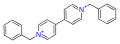 Benzyl Viologen (N blue).svg 650 × 239; 12 KB
-
 Benzyl Viologen.svg 650 × 239; 11 KB
-
 Brilliant Blue FCF(2).svg 819 × 374; 19 KB
-
 Caffeine dehydrogenase.svg 918 × 458; 41 KB
-
 Capreomycin IA.svg 1,177 × 653; 16 KB
-
 Capreomycin IB.svg 1,177 × 628; 16 KB
-
 Capreomycin.svg 2,904 × 1,649; 31 KB
-
 Carboxybiotin decarboxylase.svg 1,352 × 583; 711 KB
-
 Chloralose.svg 523 × 427; 2 KB
-
 Clindamycin skeletal.svg 1,562 × 615; 6 KB
-
 Cob(II)yrinic acid a,c-diamide reductase.svg 1,644 × 653; 118 KB
-
 Creatinase.svg 947 × 158; 15 KB
-
 Cynarine.svg 435 × 270; 32 KB
-
 D-alanine-(R)-lactate ligase.svg 571 × 207; 24 KB
-
 D-ornithine aminomutase.svg 811 × 204; 13 KB
-
 D3T (3H-1,2-dithiole-3-thiona (colour)).svg 179 × 122; 4 KB
-
 D3T (3H-1,2-dithiole-3-thiona).svg 179 × 122; 4 KB
-
 DDTHCL.svg 570 × 171; 22 KB
-
 Deacetoxycephalosporin-C synthase.svg 1,272 × 357; 44 KB
-
 Deacetylipecoside synthase.svg 1,438 × 431; 44 KB
-
 Deoxyribonuclease (pyrimidine dimer) - Steps.svg 1,126 × 543; 173 KB
-
 DGTPase.svg 1,849 × 436; 43 KB
-
 Diacylglycerol cholinephosphotransferase.svg 1,139 × 415; 49 KB
-
 DiferenceBetweenUridine-PseudouridineENG.svg 428 × 277; 23 KB
-
 DNA With cyclobutane pyrimidine dimer.svg 618 × 271; 47 KB
-
 Droperidol.svg 512 × 248; 5 KB
-
 DyCfin.svg 406 × 297; 9 KB
-
 DyT.svg 350 × 256; 9 KB
-
 Exonuclease type a.svg 704 × 672; 107 KB
-
 Exonuclease type b.svg 561 × 659; 110 KB
-
 Fluocortolone.svg 500 × 250; 25 KB
-
 Gamma-glutamil carboxilasa.svg 1,646 × 461; 36 KB
-
 Gamma-glutamyl carboxylase.svg 1,646 × 461; 36 KB
-
 Glucose-6-phosphate dehydrogenase (coenzyme-F420).svg 1,377 × 654; 55 KB
-
 GTP diphosphokinase1.svg 697 × 476; 78 KB
-
 GTP diphosphokinase2.svg 697 × 476; 73 KB
-
 Heme c.svg 334 × 431; 22 KB
-
 Heme o.svg 1,060 × 1,343; 7 KB
-
 Heparan Sulfate.svg 1,199 × 746; 43 KB
-
 HMB-PP.svg 426 × 282; 8 KB
-
 Hydroxyprogesterone caproate.svg 512 × 240; 6 KB
-
 Hygromycin A Molecular Structure.svg 1,035 × 450; 43 KB
-
 Lathosterol oxidase.svg 1,220 × 351; 37 KB
-
 LNA.svg 398 × 356; 13 KB
-
 LNASchem.svg 512 × 490; 7 KB
-
 Lobeline.svg 1,814 × 665; 6 KB
-
 Luminol chemiluminescence molecular representation.svg 4,450 × 2,694; 161 KB
-
 Lycopene ε-cyclase.svg 780 × 585; 15 KB
-
 Lycopene ε-cyclase2.svg 504 × 304; 37 KB
-
 Lysolecithin acylmutase.svg 527 × 477; 20 KB
-
 Magnesium chelatase.svg 655 × 288; 40 KB
-
 Maltose epimerase (2).svg 1,317 × 314; 31 KB
-
 Maltose epimerase.svg 1,317 × 314; 27 KB
-
 Mandelate racemase (2).svg 569 × 211; 13 KB
-
 Metabolic pathway- pyridoxal 5'-phosphate biosynthesis I v 2.0.svg 2,787 × 2,112; 125 KB
-
 Metabolic pathway- pyridoxal 5'-phosphate biosynthesis I.svg 2,411 × 2,036; 124 KB
-
 Methionine racemase.svg 1,016 × 371; 13 KB
-
 Methylmalonyl-CoA.svg 460 × 500; 60 KB
-
 Mevalonate Pathway Vector.svg 612 × 880; 94 KB
-
 Microsomal epoxide hydrolase.svg 1,381 × 364; 17 KB
-
 Morindin.svg 1,650 × 1,350; 5 KB
-
 Morpholino-monomer.svg 150 × 159; 9 KB
-
 NAD+ synthase (glutamine-hydrolysing).svg 732 × 240; 63 KB
-
 Nicotinate dehydrogenase (cytochrome).svg 384 × 177; 16 KB
-
 Nicotinate dehydrogenase.svg 542 × 244; 14 KB
-
 Nimesulide.svg 512 × 534; 5 KB
-
 Ornithine cyclodeaminase.svg 1,157 × 389; 13 KB
-
 Oxaloacetate tautomerase.svg 684 × 211; 12 KB
-
 Oxalomalate lyase (1).svg 953 × 244; 19 KB
-
 Oxalomalate lyase (2).svg 953 × 244; 18 KB
-
 Oxpheneridine.svg 700 × 400; 2 KB
-
 Pathway Atrazine degradation.svg 622 × 516; 44 KB
-
 Pheneridine.svg 700 × 390; 2 KB
-
 Piridoxina 5'-fosfato sintasa.svg 1,439 × 895; 35 KB
-
 Pivmecillinam.svg 845 × 682; 3 KB
-
 Propylene skeletal.svg 340 × 160; 665 bytes
-
 Prostaglandin-A1 Δ-isomerase.svg 363 × 485; 18 KB
-
 Prostaglandin-D synthase.svg 417 × 531; 25 KB
-
 PseudoUracil.svg 234 × 237; 6 KB
-
 Pyridoxine 5-dehydrogenase.svg 1,108 × 310; 16 KB
-
 Pyrimidodiazepine synthase.svg 1,062 × 430; 58 KB
-
 Reacción de la Glucosa 6-fosfatasa.svg 385 × 159; 24 KB
-
 Rivoflavinase.svg 1,404 × 328; 32 KB
-
 Silyl General.svg 333 × 308; 6 KB
-
 Silyl.svg 295 × 305; 4 KB
-
 Sterol synthesis pt.svg 571 × 640; 21 KB
-
 Sterol synthesis.svg 571 × 640; 21 KB
-
 Tannase.svg 1,832 × 399; 29 KB
-
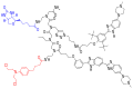
 Targaprimir 96.svg 1,568 × 832; 44 KB
-
 Targaprimir-96-CA-Biotin (1).svg 1,568 × 1,249; 57 KB
-
 Targaprimir-96-CA-Biotin (2).svg 1,574 × 1,049; 56 KB
-
 Targaprimir-96-CA-Biotin (3).svg 1,574 × 1,049; 59 KB
-
 Tenofovir disoproxil fumarate.svg 464 × 445; 23 KB
-
 Tetrahydrobiopterin tautomers.svg 530 × 704; 25 KB
-
 Thiane.svg 117 × 142; 3 KB
-
 Thiane2.svg 116 × 145; 3 KB
-
 Thiane3.svg 98 × 126; 3 KB
-
 Toluidine Blue.svg 350 × 211; 10 KB
-
 Trans-acenaphthene-1,2-diol dehydrogenase.svg 557 × 238; 17 KB
-
 Tryptophan 2'-dioxygenase.svg 651 × 288; 21 KB
-
 Tyrosine aminotransferase.svg 1,141 × 222; 27 KB
-
 Ureidoglycolate lyase.svg 915 × 172; 13 KB
-
 Uroerythrin.svg 632 × 334; 13 KB
-
 Vitamin K epoxide reductase.svg 492 × 259; 33 KB
-
 Vía del Mevalonato.svg 612 × 880; 96 KB
-
 Xanthine dehydrogenase.svg 736 × 202; 19 KB
-
 Xilose degradation I en.svg 754 × 460; 28 KB
-
 Xilose degradation I es.svg 754 × 460; 28 KB


.svg)
purine.svg)


.svg)


.svg)
.svg)


.svg)


).svg)
.svg)
_-_Steps.svg)


.svg)


.svg)


.svg)
.svg)
.svg)


arginine_beta-lactam-synthase.svg)
phenol_mutase.svg)
.svg)
.svg)
.svg)
yrinic_acid_a,c-diamide_reductase.svg)
-lactate_ligase.svg)
.svg)
.svg)
.svg)
.svg)
.svg)