Commons:Wiki Science Competition 2019/Winners
Evaluation process edit
For more information about the selection procedure please go here.
National finalists edit
In this table you can find all information about national finalists (and eventually, national winners) of WSC2019.
Click on these page to know all information related to local jurors, timelines and finalists.
Australia · Estonia · France · India · Ireland · Italy · Malaysia · New Zealand · Poland · Portugal · Russia · Spain · Switzerland · Ukraine · United Kingdom · United States ··· The rest of the World
Second international round edit
The national finalists are selected in an intermediate round focused mostly on the originality and the image quality.
- For the categories processed with Montage (people in science, microscopy images, wildlife and nature and general category), only the images with more than 6 out of 10 averaged votes are showed in this summary. All jurors gave a vote for 0.0 to 1.0 to every images.
- The other categories (non photographic media, set of images) were processed with a xls file, and a different scale was used where all the jurors gave votes 10 (best) to 1 (worst) to their first ten choices. Files that were not selected by the jurors were assigned "0.0". The average is lower than in the previous case, and the 10-12 voted choices are shown.
Similarly to Wiki Loves Monuments, international and national juries can show different taste, especially when some juries are small. For this reason it possible that some second finalists are given a higher ranking than the national winners at the international level.
Winners edit
The final winner and 4-6 runners-up were finally selected out of a final brainstorming from the finalists after the intermediate round, with some minor rearrangement based on
- the quality of their scientific description;
- the resolution and the presence of technical and scientific details;
- specific strong criticism by jurors or the academic committee (e.g. very common idea);
- use on wikimedia platforms (as of July 2020);
 Featured file
 Quality images
 Valued file
 Used on other wikimedia platforms
- excessive similarity to the winners and runners-up of the previous edition;
- a final balance of subjects and themes.
Please notice that in order to maximize the chance of every file, and due to specific taste of national and international juries, some files are categorized twice.[1]
People in Science edit
|

|
I'm a physics PhD student based at the National Australian University, Canberra, Australia. I always thought black holes were cool but distorted space-time and gravitational waves got me hooked -- so I joined the LIGO Scientific Collaboration during my final year of undergrad in 2014. I was working as an operations specialist in the control room at LIGO Hanford, one of the two U.S. based gravitational-wave detectors, when the first gravitational wave was detected in September 2015. Couple years later I left Washington state to pursue a PhD in the Down Under. After having spent less than a year in Australia my advisor sent me back to LIGO Hanford to help construct and commission the quantum squeezing system (aka "squeezer") prior to the third observing run and again during the third observing run break. I would always carry a camera around for documentation purposes. One of my hobbies happened to be street photography so unconsciously I would point my camera at people in addition to documenting the science for my thesis. On a rare day when I didn't have to work at my own corner, I followed Georgia Mansell (MIT PostDoc) and Jason Oberling (site detector engineer) into the Pre-Stabilized Laser (PSL) enclosure. It was my first time seeing the inside of one of the most guarded areas of LIGO. Inside the PSL is where the laser we use to detect gravitational waves generates. It is literally where it all begins. The system is capable of outputting 70-80 W of laser power so the room needs to be clean as dust and bugs can damage the optic coatings if they burn. While I was watching Georgia and Jason work I snapped this photo as the two were baffled by the low amount of light coupling into the new fiber coupler they just installed. Just like every other equipment, LIGO too needs to be maintained constantly. When we read about scientific discoveries on the news or hear science talks at conferences often the human factor is neglected. LIGO isn't just a giant machine that listens to black holes and neutron star collisions. It takes decades and generations of scientists to get to where it is today. Routine maintenance is also required to keep the detector(s) operational. By sharing this photo with a wider audience via the competition I hope to bring humans back into science. Science is cool, I know, but after all it's the quirky human scientists that make the science happen and inspire generations to come. |
| International runner-up | International runner-up | |||
| Weekly maintenance of the needles on the clinical chemistry module. |
The office of the paleoanthropologist Stefano Ricci at the University of Siena. |
|||


| International runner-up | International runner-up | |||
| Associate Professor in Estonian Geography Taavi Pae in his office. |
Junior researcher Andrei Kartoziia at the Samoylov Island scientific station. |
|||


| International runner-up | International runner-up | |||
| A prototype of a snake-like robot for endoscopic surgery: the i²Snake. |
Heart valve prosthesis implantation. Amosov National Institute of Cardiovascular Surgery. |
|||


Wildlife and Nature edit
|

|
I am a PhD student with La Trobe University in Melbourne, Australia, specialised in the field of seabird ecology. Amongst other researching goals I am currently working on a novel monitoring technique for the breeding population of the endangered Abbott’s booby (Papasula abbotti), which today is only known to nest on Christmas Island. I first experienced Christmas Island in 2010 as a field volunteer of a former seabird researcher from the University of Hamburg, Germany – where I studied Biology at that time. During one of multiple revisits in the years after graduating with a Master in Science, I was lucky to meet my present supervisor during one of his research field trips on Christmas Island which resulted in the opportunity to start a PhD project that focuses on one of the most charismatic seabird species and, for an ecologist, many technically interesting aspects. This photo was taken during one of the initial trials to determine if we could detect Abbott’s Booby nests from the air with an off-the-shelf drone. A ground-based survey for this species is extremely labour intensive as Abbott’s Boobies breed sparsely in mostly 30-40 m high dense rainforest canopies with thick and sometimes difficult to traverse understory due to invasive species, historical forest clearing, and climate change impacts that altered the native forest over years. The use of a drone eliminates some of these difficulties but does not come without challenges. The high trees and the often small and rare tracks in the jungle of Christmas Island for drone operations while employing line-of-sight direction as per air traffic regulations are one of them. Nevertheless, we were able test this method for a good percentage of potential Abbott’s Booby habitat and we are hoping to present the results soon. In cooperation with the University of East Anglia in Norwich, United Kingdom, we are currently working on automated detection – an important step to proceed this methodology to an efficient tool for the population assessment of the Abbott’s booby. Having searched for nests on many hundreds of photographs manually already, I still find the view from above to the dense rainforest canopies like a refreshing oasis full of clean oxygen, and personally am sometimes slightly jealous of the bird’s perspective to life. With this novel approach of drone technology applied to wildlife monitoring, I am honoured to share this view through the Wikipedia Science competition. |
| International runner-up | International runner-up | |||
| Killer whales spyhopping to locate a crabeater seal on an ice floe in Antarctica. |
Aurora in Kandalaksha, Murmansk region. |
|||


| International runner-up | International runner-up | |||
| Ringing of an Ural owl (Strix uralensis). |
Immature Phidippus jumping spider in Pennsylvania. |
|||
_ringing.jpg)

| International runner-up | International runner-up | |||
| Common Coot or Eurasian Coot (Fulica atra) scoots across the water on Chambal River. |
Caenolyda reticulata. |
|||


Microscopy images edit
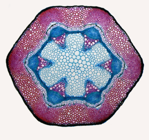
Prize motivation: Nature is life, and no message related to life is more powerful than birth. The image might be relatively know to a more specialized audience, but it is definitively attractive for the general public, thanks to the combination of the unusual topic with the mesmerizing color pattern, and it encourages to know more about reproduction in a phylum different than ours. |

|
I am a biologist by profession. I have been interested in photography since I was 12 years old. I was always interested in the world of small things, and that is why macro photography attracted me from the very beginning. The adventure with the microscope began a year or two later, when I dug out of my father's dusty and old Paul de Kruif's book "Microbe hunters." This book changed my life. Although I have always been interested in nature, reading this book directed my interests towards a completely different world, the one imperceptible to the naked eye. I wanted to discover the secrets of the microcosm. I built my own microscopes, and what I saw through them, I drew. I drew primarily for myself, like many before me. In the early eighties I took my first black-and-white photomicrograph. Due to various problems of that time (hardware shortages, financial difficulties), I abandoned microphotography for a long time, until 2009, when I bought my first digital camera. Digital technology has opened up completely new possibilities for me. Since then, I have devoted myself to this field completely. I photograph not only for myself, I also want to show to others the undeniable and often surprising beauty of the microworld. Sometimes, while we observe the world under a microscope, in addition to admiring the microcosm, we can witness certain biological processes happening here and now. We can, for example, observe the proliferation of protozoa by division or, the conjugation of the artery. With a bit of luck, we can sometimes observe the mystery of the birth of a new organism, as was the case with this winning photography. The photo was made in a combined lighting technique - dark field and polarization. It is thanks to these techniques that the photographed object owes its rich colors. The photo shows more young daphnia waiting in the mother's breeding chamber to be born, to cope with its challenges, and a little later to release the next generation. |
| International runner-up | International runner-up | |||
| Crystals, mainly sugar, in a dried Coca Cola droplet, taken with a microscope using cross polarization. |
Eye of Chrysopidae (stacking of 30 frames). |
|||


| International runner-up | International runner-up | |||
| 3D projection of a Patiria miniata bipinnaria. |
"Coffee ring" from aluminium oxide particles (150 nm) on cover glass coated with gold (thickness 20 nm). |
|||

_%D0%BD%D0%B0_%D0%BF%D0%BE%D0%B4%D0%BB%D0%BE%D0%B6%D0%BA%D0%B5_%D0%B8%D0%B7_%D0%BF%D0%BE%D0%BA%D1%80%D0%BE%D0%B2%D0%BD%D0%BE%D0%B3%D0%BE_%D1%81%D1%82%D0%B5%D0%BA%D0%BB%D0%B0,_%D0%BF%D0%BE%D0%BA%D1%80%D1%8B%D1%82%D0%BE%D0%B9_%D0%B7%D0%BE%D0%BB%D0%BE%D1%82%D0%BE%D0%BC_%D1%82%D0%BE%D0%BB%D1%89%D0%B8%D0%BD%D0%BE%D0%B9_20_%D0%BD%D0%BC.jpg)
| International runner-up | International runner-up | |||
| Escherichia coli growing on Eosin methylene blue (EMB) media with its distinctive green metallic sheen. |
Surface oxides of conductive 3D copper structures visible under the microscope as a blueish glimmer. |
|||


Non-photographic media edit
|

|
I am a professor of physics and of astronomy at UC Berkeley and my research focus is cosmology. One of the main goals of cosmology is to elucidate the origins of our Universe. We believe our Universe started with a rapid expansion called inflation, during which tiny quantum seeds were imprinted into the fabric of spacetime, leading in time to creation of galaxies, stars and planets, including the Earth. To prove this theory myself and my colleagues proposed to search for B-modes, a spiral type of polarization imprinted in the microwave sky. However, Milky Way emits dust, which is also polarized, and scientists must learn how to distinguish the two signals. This image was created by taking dust emission as measured from the Planck satellite and converting its intensity into a polarization pattern. The bright yellow band is Milky Way as seen in the microwave sky. A B-mode was imprinted into a single spot by locally rotating the dust polarization pattern by 45 degrees. A hilly landscape was added at the bottom to represent that many of the B-mode experiments are ground based, in places from Antarctica to Atacama desert in Chile. This picture is a great example of a teamwork in science and could not have been made without the input of numerous people. Special thanks to the Planck satellite team who observed and made public the data this is based on, Susan Clark for the original inspiration, Yu Feng for the data processing and Andrej Berlot for the post-processing. |
| International runner-up | International runner-up | |||
| In-depth characterization of Deinococcus radiodurans cell cycle. |
MRI self portrait. |
|||

| International runner-up | International runner-up | |||
| Relief of Tenerife processed with the QGIS geographic information system and made from a digital LiDAR elevation model. |
The growth of silver crystals on the surface of copper from a solution of silver nitrate. |
|||

| International runner-up | International runner-up | |||
| Dripping with refractive index variation. The motion is created by a difference of surface tension between miscible fluids. |
RNA polymerase (purple) with one DNA double helix strand (darker orange) and RNA (green). |
|||


General category edit
|

|
I am a technician at the Anthropology Laboratory of the Department of Physical Sciences, Earth and Environment (Research Unit in Prehistory and Anthropology) of the University of Siena and a member of the Fisiocritici Academy of Siena. Thanks to my skills in anatomical drawing, I have had the opportunity to do scientific illustrations and replicas of fossil specimens for Italian and foreign museums and magazines. I combined my artistic skills with a constant commitment in the academic field, as testified by publications in physical paleoanthropology in prestigious scientific international journals. Photography is also a very important part of my work and allowed me to do the cover images of the 2003 May issue of PNAS and the 2019 October issue of Nature - Ecology&Evolution. My research group is focusing on the reconstruction of the behavior and subsistence strategies of past human populations, and is carrying out excavations in some of the key sites of Paleolithic Italy. This activity requires the work of many specialists from different disciplines, engaged both in laboratory activities and in the field. My photographs thus become a sort of trait d'union that fixes in time research phases, studied contexts and archaeological specimens from a distant past. Photographic documentation is fundamental in every phase of our research, not only in the lab, but also during fieldworks. This, in fact, represents the unrepeatable moment in which the finds (future research data) are extracted from the sediment that sealed them. Behind every excavation there are people, stories and close collaborations between many different specialists. This is true in "ordinary" excavations but even more so in the more "special" ones, such as the prehistoric site of Grotta dei Santi (Municipality of Monte Argentario, Tuscany, central Italy), where this picture was taken. The site is in excavation by the Department of Physical Sciences, Earth and Environment of the University of Siena in collaboration with the Soprintendenza Archeologia, Belle Arti e Paesaggio per le Province di Siena, Grosseto e Arezzo. Some of the last Italian Neanderthals dwelled in this site between 50,000 and 40,000 years ago. The cave is accessible only by sea, which gives to the site a breath-taking scenery. For logistical reasons, during the fieldwork, the life of the scholars takes place almost entirely in the cave. Each archaeologist has his own tent, the base camp is installed a few meters from the excavation area, where the Neanderthal campsites were set thousands years ago. A very immersive experience! In such an excavation, coordination and cooperation become indispensable. Among the numerous institutions involved, the Fire Brigade of Grosseto, which is present in this photo, is fundamental for the invaluable logistic support they offered to the team. |
| International runner-up | International runner-up | |||
| Aerial view of the tributary of the Chusovaya river, Sverdlovsk region.. |
Telescope's tower crane at Large azimuth telescope. |
|||


| International runner-up | International runner-up | |||
| The road down from Teide volcano in Tenerife with the Milky way above it. |
Environmental disaster in Levikha Village, because of the massive sulfide underground mine. |
|||


| International runner-up | International runner-up | |||
| Meteor, satellite and aeroplane over the Paranal Observatory, Chile, with a stargazer in the foreground. |
Li-ion battery cell exploded after hitting it by hammer. |
|||


Image sets edit
|


|
I wanted to show this beauty to others. In the last century, I used a camera with color film, and I had to take a lot of frames, print photographs, most of which ended up in the trash can. Digital cameras nowadays allow you to immediately observe and capture the image from the microscope. I have created a research laboratory for children called "Microcosm", where students are free to study the topic of the "little world". For many years I have been investigating plant anatomy, making original permanent preparations. Most of my slides are hand-made. Every year, in the summer, my students and I go on botanical expeditions, studying rare plants of Siberia. On the expeditions, we also collect material for future preparations. There are several items in these photographs, all hand-made and prepared by me.
|
| International runner-up | International runner-up | |||
| Special image treatment process of the "Vase of the Warriors" of Archena. |
Close-up photographs of insect heads. |
|||


| International runner-up | International runner-up | |||
| Simulations of magnetic field and density turbulence in collisionless accretion disk plasma around supermassive black hole. |
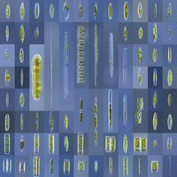
Diatoms from ecosystems in the Moscow region. Images obtained with differential interference contrast.
|
|||


| International runner-up | International runner-up | |||
| Scientists entering into cave to monitor bats in Estonia.
|
Technique of rollout or perimeter photography, reconstructing artifacts with Microsoft ICE (Image Composite Editor) |
|||


Special Q-SORT prizes edit
Special prized by the EU-funded Q-SORT consortium. Q-SORT has received funding from the European Union’s Horizon 2020 Research and Innovation Programme under Grant Agreement No. 766970 Q-SORT (H2020-FETOPEN-1-2016-2017).
| Q-SORT Prize "people in science" | Q-SORT Prize "microscopy" | |||
| Homeschooled Sofia (6 years old) is presenting her finished project on our solar system. |
Transmission electron microscope image of a tightly-focused laser beam, showing the individual crests and troughs of its electromagnetic wave. |
|||


- ↑ For example there is a new "wildlife and nature" category, but some landscapes are also considered under "general", similarly certain jury accept images in the "people in science" category where only a portion of the body is visible, so they are sometimes included in other categories as well at the international level.