File:A-Distinct-Layer-of-the-Medulla-Integrates-Sky-Compass-Signals-in-the-Brain-of-an-Insect-pone.0027855.s005.ogv
Size of this JPG preview of this OGG file: 790 × 600 pixels. Other resolutions: 316 × 240 pixels | 632 × 480 pixels | 1,011 × 768 pixels | 1,264 × 960 pixels.
Original file (Ogg Theora video file, length 8.2 s, 1,264 × 960 pixels, 5.25 Mbps, file size: 5.1 MB)
Captions
Captions
Add a one-line explanation of what this file represents
Summary edit
| Description |
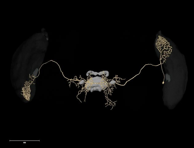
English: Movie showing a 3D model of the MeMe2 neuron (same as in Figure 4F). Optic lobe neuropils and brain areas of the central brain are shown in transparent grey. Vertical rotation. |
||
| Date | |||
| Source | Movie S5 from el Jundi B, Pfeiffer K, Homberg U (2011). "A Distinct Layer of the Medulla Integrates Sky Compass Signals in the Brain of an Insect". PLOS ONE. DOI:10.1371/journal.pone.0027855. PMID 22114712. PMC: 3218074. | ||
| Author | el Jundi B, Pfeiffer K, Homberg U | ||
| Permission (Reusing this file) |
|
||
| Provenance |
|
File history
Click on a date/time to view the file as it appeared at that time.
| Date/Time | Thumbnail | Dimensions | User | Comment | |
|---|---|---|---|---|---|
| current | 16:40, 17 November 2012 | 8.2 s, 1,264 × 960 (5.1 MB) | Open Access Media Importer Bot (talk | contribs) | Automatically uploaded media file from Open Access source. Please report problems or suggestions here. |
You cannot overwrite this file.
File usage on Commons
There are no pages that use this file.