File:A-Spinal-Cord-Window-Chamber-Model-for-In-Vivo-Longitudinal-Multimodal-Optical-and-Acoustic-Imaging-pone.0058081.s005.ogv
Size of this JPG preview of this OGG file: 800 × 537 pixels. Other resolutions: 320 × 215 pixels | 640 × 429 pixels | 912 × 612 pixels.
Original file (Ogg Theora video file, length 4 min 9 s, 912 × 612 pixels, 206 kbps, file size: 6.12 MB)
Captions
Captions
Add a one-line explanation of what this file represents
Summary edit
| Description |
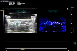
English: Spinal cord O2 saturation monitoring by photoacoustic imaging. Two-dimensional cross-section of the spinal cord within the window chamber. Ultrasound structural image (left) shows the outline of the window chamber as well as the artificial dura that cover the spinal cord. The rectangle indicates the region where photoacoustic image was acquired, and the circular region of interest indicates the area that photoacoustic signal intensity was measured. Photoacoustic image (right) displays the spinal cord vasculature. Color bar indicates the relative oxygenation level of the vasculature, and the scale bar illustrates the depth of imaging from the transducer head. The animal’s anaesthetic mixture was shifted from 100% to 7% oxygen for 1 minute, which corresponds to the frame 58 to 103 (out of total 248 frames acquired) in this video. |
||
| Date | |||
| Source | Video S3 from Figley S, Chen Y, Maeda A, Conroy L, McMullen J, Silver J, Stapleton S, Vitkin A, Lindsay P, Burrell K, Zadeh G, Fehlings M, DaCosta R (2013). "A Spinal Cord Window Chamber Model for In Vivo Longitudinal Multimodal Optical and Acoustic Imaging in a Murine Model". PLOS ONE. DOI:10.1371/journal.pone.0058081. PMID 23388716. PMC: 3597636. | ||
| Author | Figley S, Chen Y, Maeda A, Conroy L, McMullen J, Silver J, Stapleton S, Vitkin A, Lindsay P, Burrell K, Zadeh G, Fehlings M, DaCosta R | ||
| Permission (Reusing this file) |
|
||
| Provenance |
|
File history
Click on a date/time to view the file as it appeared at that time.
| Date/Time | Thumbnail | Dimensions | User | Comment | |
|---|---|---|---|---|---|
| current | 03:05, 22 March 2013 | 4 min 9 s, 912 × 612 (6.12 MB) | Open Access Media Importer Bot (talk | contribs) | Automatically uploaded media file from Open Access source. Please report problems or suggestions here. |
You cannot overwrite this file.
File usage on Commons
There are no pages that use this file.