File:Anemonia sargasensis.jpg

Size of this preview: 539 × 599 pixels. Other resolutions: 216 × 240 pixels | 432 × 480 pixels | 691 × 768 pixels | 921 × 1,024 pixels | 1,512 × 1,681 pixels.
Original file (1,512 × 1,681 pixels, file size: 2.78 MB, MIME type: image/jpeg)
Captions
Captions
Add a one-line explanation of what this file represents
Summary edit
| Description |
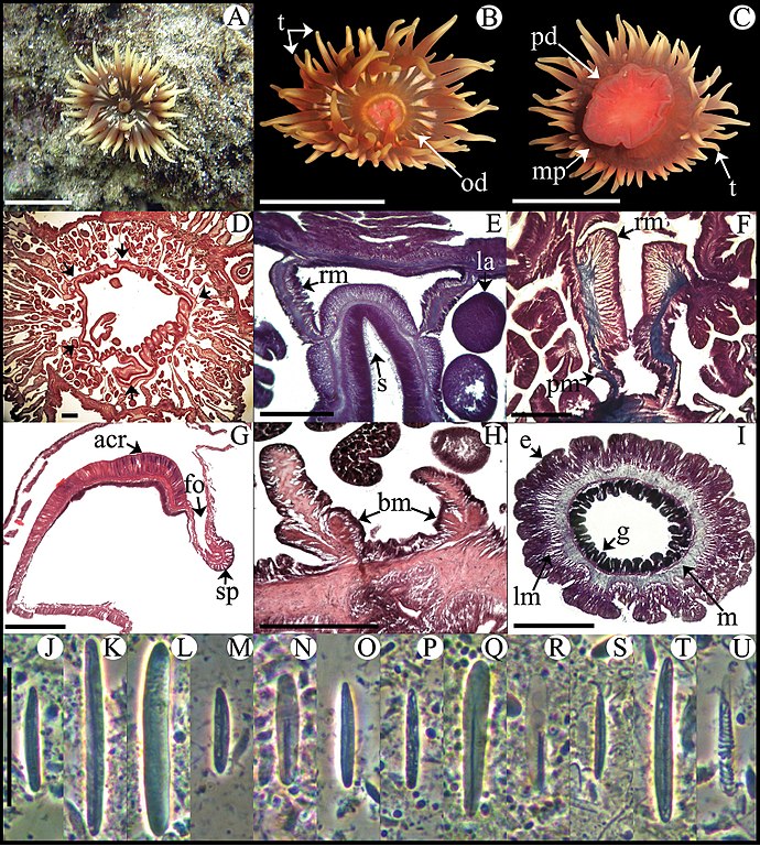
Español: Anemonia sargassensis. A Espécimen vivo en su hábitat natural B Vista oral C Vista de disco pedal D Sección transversal de la columna distal mostrando mesenterios; las flechas indican sifonoglifos E Detalle de sección transversal de la columna distal mostrando un sifonoglifo F Detalle de músculos retractor y parieto-basilar G Sección longitudinal del margen mostrando acrorhagi y músculo marginal del esfinter H Sección longitudinal de la base mostrando músculos basilares I Sección transversal de tentáculo J–U Cnidae.– acrorhagi: J pequeño basitrico K basitrico L holotrico; actinofaringe: M pequeño basitrico N microbasic p-mastigoforo; columna: O basitrico; filamentos: P basitrico Q microbasic b-mastigoforo R microbasic p-mastigoforo; tentáculo: S pequeño basitrico T basitrico U espirocisto. Abreviaturas.– acr: acrorhagi, bm: músculo basilar, e: epidermis, fo: fosa, g: gastrodermis, la: larva, lm: músculo longitudinal, m: mesoglea, mp: proyección marginal, od: disco oral, pd: disco pedal, pm: músculo parietobasilar, rm: músculo retractor, s: sifonoglifo, sp: esfinter, t: tentáculo. Barras de escala: A–C: 10 mm; D–I: 200 μm; J–U: 25 μm. English: Anemonia sargassensis. A Live specimen in natural habitat B Oral view C Pedal disc view D Cross section through distal column showing mesenteries; arrows indicate siphonoglyphs E Detail of cross section through distal column showing a siphonoglyph F Detail of retractor and parietobasilar muscles G Longitudinal section through margin showing acrorhagi and marginal sphincter muscle H Longitudinal section through base showing basilar muscles I Cross section through tentacle J–U Cnidae.– acrorhagi: J small basitrich K basitrich L holotrich; actinopharynx: M small basitrich N microbasic p-mastigophore; column: O basitrich; filaments: P basitrich Q microbasic b-mastigophore R microbasic p-mastigophore; tentacle: S small basitrich T basitrich U spirocyst. Abbreviations.– acr: acrorhagi, bm: basilar muscle, e: epidermis, fo: fosse, g: gastrodermis, la: larvae, lm: longitudinal muscle, m: mesoglea, mp: marginal projection, od: oral disc, pd: pedal disc, pm: parietobasilar muscle, rm: retractor muscle, s: siphonoglyph, sp: sphincter, t: tentacle. Scale bars: A–C: 10 mm; D–I: 200 μm; J–U: 25 μm. |
| Date | |
| Source |
https://zookeys.pensoft.net/articles.php?id=3319&display_type=list&element_type=9 |
| Author | Ricardo González-Muñoz, Nuno Simões, José Luis Tello-Musi, Estefanía Rodríguez |
Licensing edit
This file is licensed under the Creative Commons Attribution 3.0 Unported license.
- You are free:
- to share – to copy, distribute and transmit the work
- to remix – to adapt the work
- Under the following conditions:
- attribution – You must give appropriate credit, provide a link to the license, and indicate if changes were made. You may do so in any reasonable manner, but not in any way that suggests the licensor endorses you or your use.
File history
Click on a date/time to view the file as it appeared at that time.
| Date/Time | Thumbnail | Dimensions | User | Comment | |
|---|---|---|---|---|---|
| current | 11:12, 5 December 2015 | | 1,512 × 1,681 (2.78 MB) | Josuevg (talk | contribs) | User created page with UploadWizard |
You cannot overwrite this file.
File usage on Commons
There are no pages that use this file.