File:Blindness Malformation 2.jpg

Original file (689 × 838 pixels, file size: 83 KB, MIME type: image/jpeg)
Captions
Captions
Summary edit
| Description |
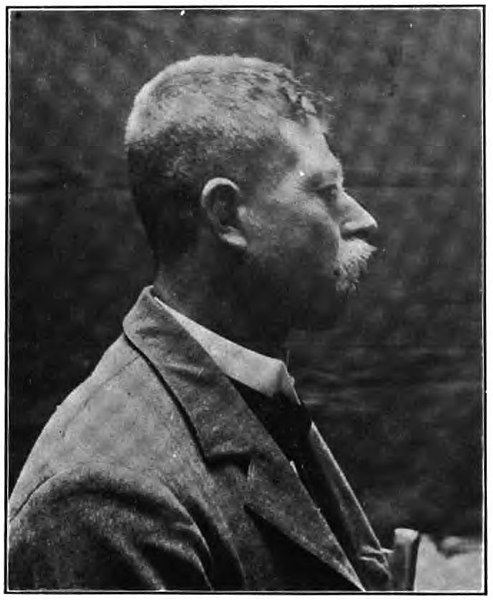
English: CASE II.-The patient, who was born in Germany, was a thirty-five-
year-old farmer. He stated that he had always had a curiously shaped skull. He had been free from all disease until he was ten years old, at which time he had had a series of spasms. These convulsions were associated with a permanent divergence of the eyes and a persistent in- different vision which was more pronounced in the left eye. Three weeks before I saw him, he noticed that the sight of his good eye began to fail, this failure being associated at times with deeply seated orbital pains on the same side. His habits, he said, were good, and there were not any signs of gross hereditary or acquired disease. No other mem- bers of his family “for three generations back had gone blind.” His parents were not blood relations. Vision with the right eye was reduced to an incorrectible one-eighth of normal in an excentrically placed field, with its fixation-point situ- ated far up and in. Color perception for green, red, blue and yellow was lost. Vision with the left eye was almost gone, there being but one small area of doubtful at times light-perception situated in an extreme temporal field as the last remnant of sensory functioning. Intraocular tension in each eye was normal. The pupil of the left eye, which was round, was about two millimeters larger than the similarly shaped one of the right eye. The right iris responded fairly well to light-stimulus and accommodative efforts, giving rise to rather prompt consensual reactions of the iris of the almost blind left eye during both of these impulses. The left iris was almost immobile to light-stimulus thrown upon its retina, but responded feebly to forced movement for supposed accommodation, and gave quite prompt consensual reaction to the iris of the less affected organ. Gross downward convergence of the two eyes, by having the patient endeavor to look at his nose tip, rapidly brought the pupillary areas down to one millimeter each in size. In spite of a left divergence of about thirty degrees out and slightly down, the exterior muscles of the two organs seemed to enjoy good movement. An almost constant lateral nystagmus that increased upon attempts at near fixation was a prominent symptom. The patient's eye-grounds were characteristic of consecutive atrophy, that of the right eye showing evidences of a recent optic neuritis of postocular type. Although not hoping for any permanency of result, I gave the patient the benefit of therapeutically driving more blood through the half- starved and degenerating neural tissues of the affected optic nerves. This was done by the internal administration of large and frequently repeated doses of strychnia, resulting in a temporary betterment." 2 I am under obligations to Dr. Clarence Van Epps, one of my Residents in both institutions, for presentation of the copy of the photograph of the first subject taken by Mr. James F. Wood, of Philadelphia; to Dr. Frederick C. Krause, one of my former assistants, and now Assistant Ophthalmic Surgeon to St. Christopher's Hospital, in Philadelphia, for photographing the second case; to Dr. William L. Zuill, one of the Assistant Surgeons at Wills' Hospital, for the craniometric measurements of the second case; and to Dr. Frank R. Harrison, of East Liverpool, Ohio, for securing the photograph of the third case. 3 Individuals from two races have been purposely used in the elucidation of this phase of the subject in order to obtain exceptionally broad standpoints of observation. 1 During a portion of my studies of this case the patient attended the public clinic of my friend, Dr. George C. Harlan, at the Pennsylvania Hospital. Dr. Harlan's findings and results of treatment coincided with my own. |
| Date | |
| Source | "Blindness From Congenital Malformation of the Skull", Proceedings of the American Philosophical Society v. 41 |
| Author | Charles A. Oliver, A.M., M.D. |
Licensing edit
This work is in the public domain in the United States because it was published (or registered with the U.S. Copyright Office) before January 1, 1929.
Public domain works must be out of copyright in both the United States and in the source country of the work in order to be hosted on the Commons. If the work is not a U.S. work, the file must have an additional copyright tag indicating the copyright status in the source country.
Note: This tag should not be used for sound recordings. |
File history
Click on a date/time to view the file as it appeared at that time.
| Date/Time | Thumbnail | Dimensions | User | Comment | |
|---|---|---|---|---|---|
| current | 21:06, 4 October 2022 | | 689 × 838 (83 KB) | Ted Shackelford (talk | contribs) | Uploaded a work by Charles A. Oliver, A.M., M.D. from "Blindness From Congenital Malformation of the Skull", Proceedings of the American Philosophical Society v. 41 with UploadWizard |
You cannot overwrite this file.
File usage on Commons
There are no pages that use this file.