File:Broca’s area and adjacent cortex.png
Size of this preview: 774 × 600 pixels. Other resolutions: 310 × 240 pixels | 619 × 480 pixels | 991 × 768 pixels | 1,054 × 817 pixels.
Original file (1,054 × 817 pixels, file size: 269 KB, MIME type: image/png)
Captions
Captions
Add a one-line explanation of what this file represents
Summary edit
| Description |
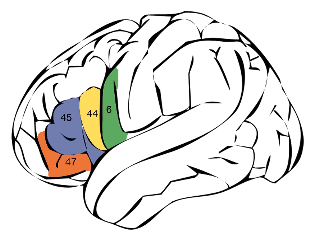
English: Schematic drawing of the lateral view of the left hemisphere and the position of the classic Broca’s area defined as encompassing Brodmann’s areas (BA) 44 (yellow) and 45 (blue) and adjacent cortex in BA 47 (orange) and ventral BA 6 (green). |
| Date | First published online: 28 June 2014 |
| Source | "Task-induced brain activity in aphasic stroke patients: what is driving recovery?" Fatemeh Geranmayeh, Sonia L. E. Brownsett, Richard J. S. Wise. Brain 2014 Oct 28;137(Pt 10):2632-48. Epub 2014 Jun 28. DOI: https://dx.doi.org/10.1093/brain/awu163 |
| Author | Fatemeh Geranmayeh, Sonia L. E. Brownsett, Richard J. S. Wise |
Licensing edit
This file is licensed under the Creative Commons Attribution 3.0 Unported license.
- You are free:
- to share – to copy, distribute and transmit the work
- to remix – to adapt the work
- Under the following conditions:
- attribution – You must give appropriate credit, provide a link to the license, and indicate if changes were made. You may do so in any reasonable manner, but not in any way that suggests the licensor endorses you or your use.
File history
Click on a date/time to view the file as it appeared at that time.
| Date/Time | Thumbnail | Dimensions | User | Comment | |
|---|---|---|---|---|---|
| current | 23:52, 2 February 2015 | | 1,054 × 817 (269 KB) | Was a bee (talk | contribs) | {{Information |Description={{en|Schematic drawing of the lateral view of the left hemisphere and the position of the classic Broca’s area defined as encompassing Brodmann’s areas (BA) 44 (yellow) and 45 (blue) and adjacent cortex in BA 47 (orange)... |
You cannot overwrite this file.
File usage on Commons
The following page uses this file: