File:Chlamydomonas TEM 06.jpg
Size of this preview: 751 × 600 pixels. Other resolutions: 301 × 240 pixels | 601 × 480 pixels | 961 × 768 pixels | 1,280 × 1,023 pixels | 1,800 × 1,438 pixels.
Original file (1,800 × 1,438 pixels, file size: 774 KB, MIME type: image/jpeg)
Captions
Captions
Add a one-line explanation of what this file represents
| Description |
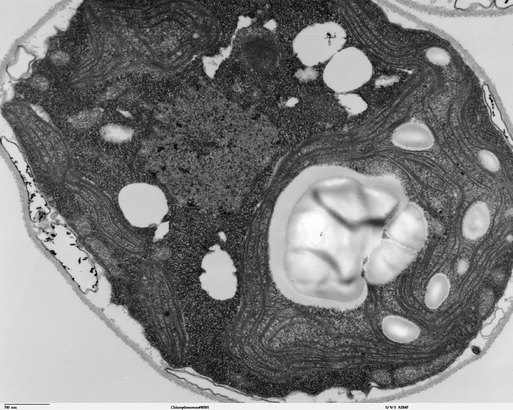
Transmission electron microscope image, showing an example of green algae (Chlorophyta). Chlamydomanas reinhardtii is a unicellular flagellate used as a model system in molecular genetics work and flagellar motility studies. This image of a thin section through a whole Chlamydomonas, shows the nucleus, chloroplast, starch grains, vacuoles, and the cell wall. |
| Date | |
| Source | Source and public domain notice at http://remf.dartmouth.edu/imagesindex.html |
| Author | Dartmouth Electron Microscope Facility, Dartmouth College |
| Permission (Reusing this file) |
Released into the public domain |
| This work has been released into the public domain by its author, Dartmouth Electron Microscope Facility, Dartmouth College. This applies worldwide. In some countries this may not be legally possible; if so: Dartmouth Electron Microscope Facility, Dartmouth College grants anyone the right to use this work for any purpose, without any conditions, unless such conditions are required by law.
|
File history
Click on a date/time to view the file as it appeared at that time.
| Date/Time | Thumbnail | Dimensions | User | Comment | |
|---|---|---|---|---|---|
| current | 07:09, 21 September 2007 | | 1,800 × 1,438 (774 KB) | Neil916 (talk | contribs) | {{Information |Description= Transmission electron microscope image, showing an example of green algae (Chlorophyta). <br><br>''Chlamydomanas reinhardtii'' is a unicellular flagellate used as a model system in molecular genetics work and flagellar motilit |
You cannot overwrite this file.
File usage on Commons
There are no pages that use this file.