File:Chromatin nucleofilaments (detail).png
Size of this preview: 800 × 480 pixels. Other resolution: 320 × 192 pixels.
Original file (1,000 × 600 pixels, file size: 502 KB, MIME type: image/png)
Captions
Captions
Add a one-line explanation of what this file represents
Summary edit
.png&action=edit§ion=1)
| Description |
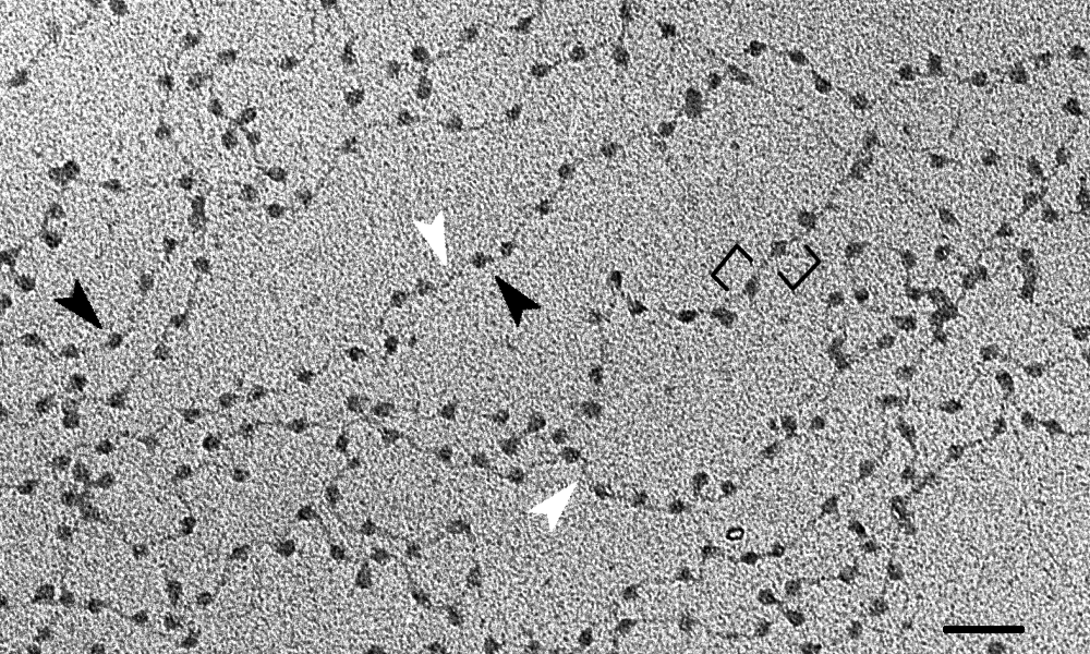
English: Electron micrograph of decondensed chromatin from chicken erythrocytes. Typical nucleofilaments (11-nm chromatin fibers, aka ”beads-on-a-string”) are seen. Black brackets highlight individual nucleosomes; black arrowheads point to nucleosome core particles (the beads), white arrowheads to linker DNA (the string). Scale bar: 50 nm. |
| Date | (UTC) |
| Source | |
| Author |
|
| This is a retouched picture, which means that it has been digitally altered from its original version. Modifications: Cropping, addition of scale bar and arrowheads to highlight interesting features. The original can be viewed here: Chromatin nucleofilaments.png:
|
Licensing edit
.png&action=edit§ion=2)
This file is licensed under the Creative Commons Attribution-Share Alike 3.0 Unported license.
- You are free:
- to share – to copy, distribute and transmit the work
- to remix – to adapt the work
- Under the following conditions:
- attribution – You must give appropriate credit, provide a link to the license, and indicate if changes were made. You may do so in any reasonable manner, but not in any way that suggests the licensor endorses you or your use.
- share alike – If you remix, transform, or build upon the material, you must distribute your contributions under the same or compatible license as the original.
Original upload log edit
.png&action=edit§ion=3)
This image is a derivative work of the following images:
- File:Chromatin_nucleofilaments.png licensed with Cc-pd
- 2011-10-14T13:20:14Z Gouttegd 3948x2773 (8638067 Bytes) {{Information |Description ={{en|1=Electron micrograph of decondensed chromatin from chicken erythrocytes. Pixel size: 0.73 nm.}} |Source =[http://www.cellimagelibrary.org/images/709 Cell Image Library 709] |Author
Uploaded with derivativeFX
File history
Click on a date/time to view the file as it appeared at that time.
| Date/Time | Thumbnail | Dimensions | User | Comment | |
|---|---|---|---|---|---|
| current | 14:34, 25 July 2015 | | 1,000 × 600 (502 KB) | Cmdrjameson (talk | contribs) | Compressed with pngout. Reduced by 169kB (25% decrease). |
| 14:04, 14 October 2011 |  | 1,000 × 600 (672 KB) | Gouttegd (talk | contribs) | == {{int:filedesc}} == {{Information |Description={{en|1=Electron micrograph of decondensed chromatin from chicken erythrocytes. Typical nucleofilaments (11-nm chromatin fibers, aka ”beads-on-a-string”) are seen. Black brackets highlight individual [[ |
You cannot overwrite this file.
File usage on Commons
The following 2 pages use this file:
File usage on other wikis
The following other wikis use this file:
- Usage on es.wikipedia.org
- Usage on fr.wikipedia.org
- Usage on uk.wikipedia.org
.png&oldid=858098776)