File:Cicadidae (10.3897-zookeys.776.26966) Figure 1.jpg

Size of this preview: 466 × 600 pixels. Other resolutions: 186 × 240 pixels | 373 × 480 pixels | 597 × 768 pixels | 795 × 1,024 pixels | 1,344 × 1,730 pixels.
Original file (1,344 × 1,730 pixels, file size: 1,013 KB, MIME type: image/jpeg)
Captions
Captions
Add a one-line explanation of what this file represents
Summary edit
_Figure_1.jpg&action=edit§ion=1)
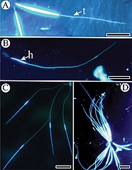
| Description | Figure 1; Epifluorescent microscope images of spermatozoa stained with Hoechst 33258. A Spermatozoon of Subpsaltria yangi with a head and a tail (t) B Spermatozoon of Platypleura kaempferi with a short head (h) and an elongated tail C Slender spermatozoa of Karenia caelatata with a head and a tail D Spermatozoa of S. yangi aggregated into bundles. Scale bars: 20 μm. |
| Date | |
| Source | https://zookeys.pensoft.net/article/26966/list/2/ (license) |
| Author | Cui B, Wei C (2018) Ultrastructure of spermatozoa in three cicada species from China (Hemiptera, Cicadomorpha, Cicadidae). ZooKeys 776: 61-80. https://doi.org/10.3897/zookeys.776.26966 |
| Permission (Reusing this file) |
This file is licensed under the Creative Commons Attribution 4.0 International license.
|
File history
Click on a date/time to view the file as it appeared at that time.
| Date/Time | Thumbnail | Dimensions | User | Comment | |
|---|---|---|---|---|---|
| current | 08:36, 19 March 2020 | | 1,344 × 1,730 (1,013 KB) | Christian Ferrer (talk | contribs) | GWToolset: Creating mediafile for Christian Ferrer. |
You cannot overwrite this file.
File usage on Commons
There are no pages that use this file.
_Figure_1.jpg&oldid=860811131)