File:Clarias gariepinus (10.3897-zoologia.37.e51603) Figures 13–18.jpg

Size of this preview: 671 × 599 pixels. Other resolutions: 269 × 240 pixels | 537 × 480 pixels | 860 × 768 pixels | 1,147 × 1,024 pixels | 1,985 × 1,773 pixels.
Original file (1,985 × 1,773 pixels, file size: 3.64 MB, MIME type: image/jpeg)
Captions
Captions
Add a one-line explanation of what this file represents
Summary edit
_Figures_13%E2%80%9318.jpg&action=edit§ion=1)
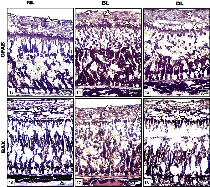
| Description | Figures 13–18; Photomicrograph of sagittal histological sections of retina of Clarias gariepinus: (13–15) showing GFAP immunostaining: (13) control showing decreased GFAP immunohistochemistry; (14)exposure to bright light showing increased immunohistochemical reaction; (15) dim light exposure showing comparatively decreased immune reaction compared to bright light;(16–18) showing BAX immunostaining. Strong reaction appeared in different retinal layers of BL and DL retina more than in normal retina. (NL) Normal light, (BL) bright light, (DL) dim light. |
| Date | |
| Source | https://zoologia.pensoft.net/article/51603/list/2/ (license) |
| Author | Sabry DA, El-Badry D (2020) Altered retina and cornea of Clarias gariepinus (Siluriformes: Clariidae) under the effect of bright and dim lights. Zoologia 37: 1-11. https://doi.org/10.3897/zoologia.37.e51603 |
| Permission (Reusing this file) |
This file is licensed under the Creative Commons Attribution 4.0 International license.
|
File history
Click on a date/time to view the file as it appeared at that time.
| Date/Time | Thumbnail | Dimensions | User | Comment | |
|---|---|---|---|---|---|
| current | 13:50, 30 December 2020 | | 1,985 × 1,773 (3.64 MB) | Christian Ferrer (talk | contribs) | GWToolset: Creating mediafile for Christian Ferrer. |
You cannot overwrite this file.
File usage on Commons
There are no pages that use this file.
_Figures_13–18.jpg&oldid=861696019)