File:Detection of lymphangiogenesis by whole-mount staining LYVE1 (red) and CD31 (green) at different time points (24 week) after vascular transplantation..png

Size of this preview: 800 × 420 pixels. Other resolutions: 320 × 168 pixels | 640 × 336 pixels | 1,024 × 537 pixels | 1,280 × 671 pixels | 3,199 × 1,678 pixels.
Original file (3,199 × 1,678 pixels, file size: 2.5 MB, MIME type: image/png)
Captions
Captions
Add a one-line explanation of what this file represents
Summary edit
_and_CD31_(green)_at_different_time_points_(24_week)_after_vascular_transplantation..png&action=edit§ion=1)
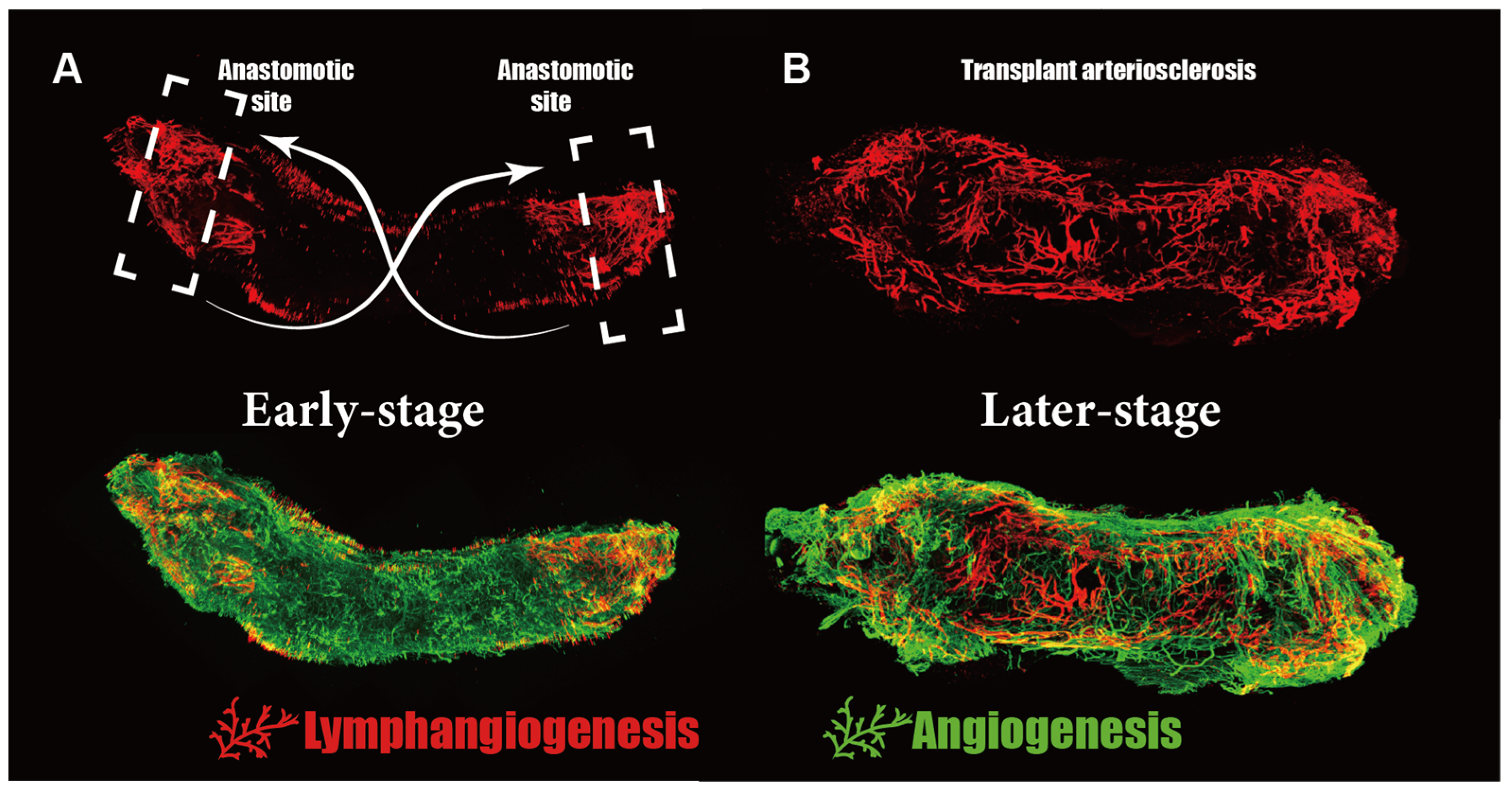
| Description | Figure 4. Detection of lymphangiogenesis by whole-mount staining LYVE1 (red) and CD31 (green) at different time points (2/4 week) after vascular transplantation. (A) The lymphangiogenesis visibly occurred at anastomotic sites and gradually spread towards the central part. (B) Until the fourth week post-transplantation, lymphatic vessels had already spread throughout the allograft vessels. Interestingly, in both time points, parts close to the anastomotic sites possessed the highest density and largest lumen area of lymphatics. |
| Date | |
| Source | https://www.mdpi.com/cells/cells-11-04056/article_deploy/html/images/cells-11-04056-g004.png https://doi.org/10.3390/cells11244056 The Impact of Stem/Progenitor Cells on Lymphangiogenesis in Vascular Disease. Cells 2022, 11, 4056. |
| Author | Mou, R.; Chen, K.; Zhu, P.; Xu, Q.; Ma, L. |

|
This file, which was originally posted to an external website, has not yet been reviewed by an administrator or reviewer to confirm that the above license is valid. See Category:License review needed for further instructions.
|
Copyright
© 2022 by the authors. Licensee MDPI, Basel, Switzerland. This article is an open access article distributed under the terms and conditions of the Creative Commons Attribution (CC BY) license (https://creativecommons.org/licenses/by/4.0/).
Licensing edit
_and_CD31_(green)_at_different_time_points_(24_week)_after_vascular_transplantation..png&action=edit§ion=2)
This file is licensed under the Creative Commons Attribution 4.0 International license.
- You are free:
- to share – to copy, distribute and transmit the work
- to remix – to adapt the work
- Under the following conditions:
- attribution – You must give appropriate credit, provide a link to the license, and indicate if changes were made. You may do so in any reasonable manner, but not in any way that suggests the licensor endorses you or your use.
File history
Click on a date/time to view the file as it appeared at that time.
| Date/Time | Thumbnail | Dimensions | User | Comment | |
|---|---|---|---|---|---|
| current | 14:14, 26 April 2024 | | 3,199 × 1,678 (2.5 MB) | Rasbak (talk | contribs) | {{Information |description=Figure 4. Detection of lymphangiogenesis by whole-mount staining LYVE1 (red) and CD31 (green) at different time points (2/4 week) after vascular transplantation. (A) The lymphangiogenesis visibly occurred at anastomotic sites and gradually spread towards the central part. (B) Until the fourth week post-transplantation, lymphatic vessels had already spread throughout the allograft vessels. Interestingly, in both time points, parts close to the anastomotic sites posse... |
You cannot overwrite this file.
File usage on Commons
There are no pages that use this file.
_and_CD31_(green)_at_different_time_points_(24_week)_after_vascular_transplantation..png&oldid=871508681)