File:Effect of TBI on Parkinsonian markers in mouse brains Impellizzeri et al (2016) FrontNeurosci 10-458.jpg

Size of this preview: 469 × 599 pixels. Other resolutions: 188 × 240 pixels | 376 × 480 pixels | 601 × 768 pixels | 1,167 × 1,491 pixels.
Original file (1,167 × 1,491 pixels, file size: 567 KB, MIME type: image/jpeg)
Captions
Captions
Effect of chronic TBI on Parkinsonian markers in mice
Summary edit
_FrontNeurosci_10-458.jpg&action=edit§ion=1)
| Description |
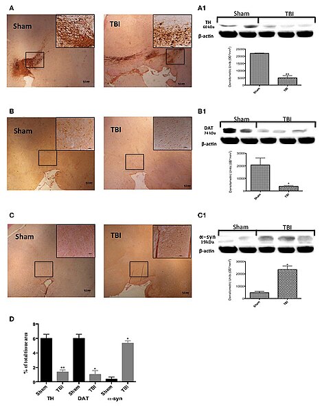
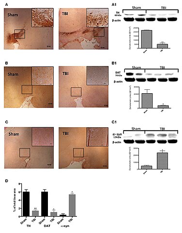
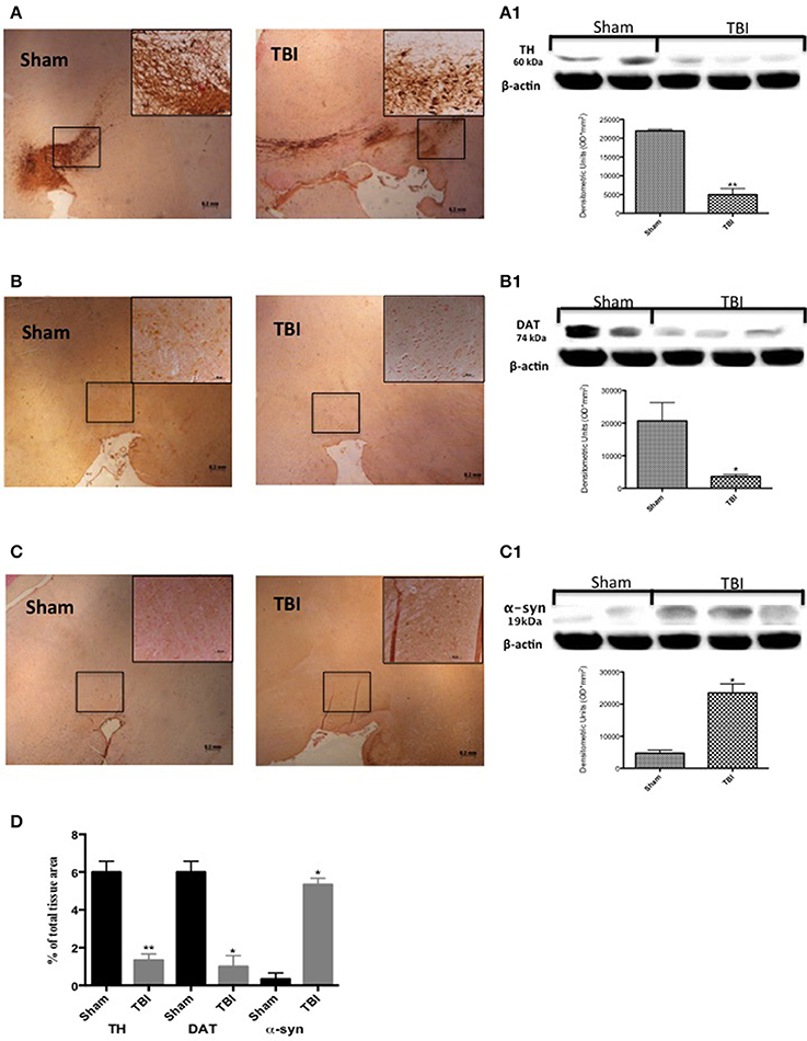
English: Original figure caption: Effect of chronic TBI on Parkinsonian markers. Midbrain was stained with antibodies against tyrosine hydroxylase (A), dopamine transporter (B) and α–synuclein (C). Immunohistochemical analysis of midbrain obtained from mice subjected to TBI revealed a positive staining for TH, DAT, and α-syn [TBI panels TBI (A–C) respectively; see densitometric analysis, D] compared with sham-operated mice [Sham panels TBI (A–C) respectively; see densitometric analysis, D]. Data are expressed as a percentage of total tissue area and are means ± SE of 5 mice/group. **P < 0.005 vs. Sham; *P < 0.05 vs. sham (Student's t-test). Western blot analysis confirmed our data (A1–C1 respectively). Each data are expressed as Mean ± SEM from N = 5 mice/group. *P < 0.01 vs. sham, **P < 0.005 vs. sham. Deutsch: Effekt chronischer Hirnschädigung infolge eines Schädel-Hirn-Traumas (SHT) auf Parkinson-Marker bei Labormäusen. Das Mittelhirn wurde eingefärbt mithilfe von Antikörpern gegen Tyrosinhydroxylase (TH) (A), Dopamintransporter (DAT) (B) und α–Synuclein (α-syn) (C). Die untersuchten histologischen Präparate stammen von Mäusen, denen im Zuge einer Schädeloperation ein SHT mittels kontrollierter kortikaler Kontusion (engl. controlled cortical impact, CCI) beigebracht wurde (Beispiele: Teilabbildungen A–C rechte Seite, „TBI“) sowie von der chirurgischen Kontrollgruppe (engl. sham), die ebenfalls einer Schädeloperation unterzogen wurden, jedoch ohne CCI (Beispiele: Teilabbildungen A–C linke Seite, „Sham“). Die Hirnpräparate der SHT-Gruppe zeigten bei den densitometrischen Messungen der eingefärbten Bereiche eine deutlich geringere Dichte von TH und DAT und eine höhere Dichte von α-syn im Vergleich zu denen der Kontrollgruppe (vgl. Teilabbildung D; angegeben sind Mittelwerte ± Standardfehler von jeweils 5 Mäusen; **P < 0.005 vs. Sham; *P < 0.05 vs. Sham (t-test)). Western-Blot-Analysen bestätigten die densitometrischen Werte (Teilabildungen A1–C1; angegeben sind Mittelwerte ± Standardfehler des Mittelwertes von jeweils 5 Mäusen; *P < 0.01 vs. Sham, **P < 0.005 vs. Sham). |
| Date | (publication date) |
| Source | Fig. 1 in: Traumatic brain injury leads to development of Parkinson’s Disease related pathology in mice. Frontiers in Neuroscience 10:458, doi:10.3389/fnins.2016.00458 |
| Author | Daniela Impellizzeri, Michela Campolo, Giuseppe Bruschetta, Rosalia Crupi, Marika Cordaro, Irene Paterniti, Salvatore Cuzzocrea, Emanuela Esposito |
| Permission (Reusing this file) |
This image was published in Frontiers in Neuroscience journal. On the website that contains the HTML version of the respective article (see DOI link above) it is stated that it “is an open-access article distributed under the terms of the Creative Commons Attribution License (CC BY). The use, distribution or reproduction in other forums is permitted, provided the original author(s) or licensor are credited and that the original publication in this journal is cited, in accordance with accepted academic practice.” |
Licensing edit
_FrontNeurosci_10-458.jpg&action=edit§ion=2)
This file is licensed under the Creative Commons Attribution 4.0 International license.
- You are free:
- to share – to copy, distribute and transmit the work
- to remix – to adapt the work
- Under the following conditions:
- attribution – You must give appropriate credit, provide a link to the license, and indicate if changes were made. You may do so in any reasonable manner, but not in any way that suggests the licensor endorses you or your use.
File history
Click on a date/time to view the file as it appeared at that time.
| Date/Time | Thumbnail | Dimensions | User | Comment | |
|---|---|---|---|---|---|
| current | 18:51, 13 April 2019 | | 1,167 × 1,491 (567 KB) | Gretarsson (talk | contribs) | {{Information |description ={{en|1=Original figure caption: '' '''Effect of chronic TBI on Parkinsonian markers.''' Midbrain was stained with antibodies against tyrosine hydroxylase (A), dopamine transporter (B) and α–synuclein (C). Immunohistochemical analysis of midbrain obtained from mice subjected to TBI revealed a positive staining for TH, DAT, and α-syn [TBI panels TBI (A–C) respectively; see densitometric analysis, D] compared with sham-operated mice [Sham panels TBI (A–C) respective... |
You cannot overwrite this file.
File usage on Commons
There are no pages that use this file.
File usage on other wikis
The following other wikis use this file:
- Usage on de.wikipedia.org
_FrontNeurosci_10-458.jpg&oldid=512341928)