File:Endoscopic-Gold-Fiducial-Marker-Placement-into-the-Bladder-Wall-to-Optimize-Radiotherapy-Targeting-pone.0089754.s001.ogv
Size of this JPG preview of this OGG file: 800 × 450 pixels. Other resolutions: 320 × 180 pixels | 640 × 360 pixels | 960 × 540 pixels.
Original file (Ogg Theora video file, length 2 min 50 s, 960 × 540 pixels, 128 kbps, file size: 2.58 MB)
Captions
Captions
Add a one-line explanation of what this file represents
Summary edit
| Description |
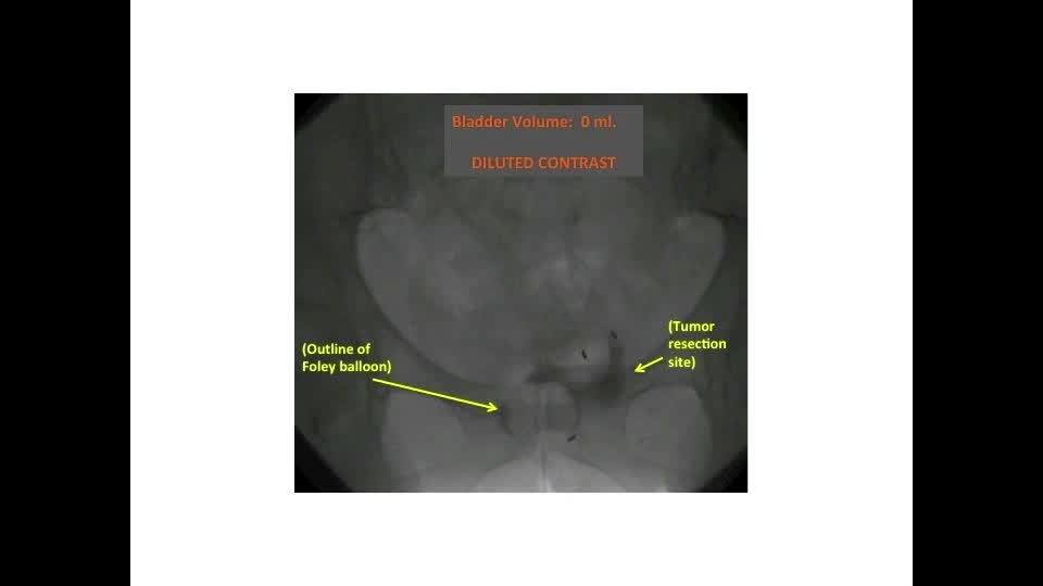
English: Three markers were placed to mark the tumor site of a patient with unifocal T2 disease. Immediately after placement, under Fluoroscopy, the empty bladder was filled and then emptied (via a urethral catheter) with dilute contrast in serial increments of 60 ml. (filling: 0 ml. to 240 ml, followed by emptying: 240 ml to 0 ml.) A Fluoroscopic image of bladder was recorded at each volume. So that the location of all markers could be easily compared relative to one another and to bony landmarks, the position of the Fluoroscopy unit and patient were fixed in space throughout imaging, so that the only source of movement was the patient's bladder and GI motility. Comparisons of images of the fiducial markers within the bladder while the latter was filled with an equal volume of either saline versus diluted contrast confirms that the markers move with the bladder-wall as the bladder expands and contracts during filling and emptying. Also, rapid serial images of the bladder were captured while holding intravesical volume constant. These images show how both the abdominal wall, intestines and the bladder-wall move in 3D with visceral peristalsis and inspiration. |
|||
| Date | ||||
| Source | Video S1 from Garcia M, Gottschalk A, Brajtbord J, Konety B, Meng M, Roach M, Carroll P (2014). "Endoscopic Gold Fiducial Marker Placement into the Bladder Wall to Optimize Radiotherapy Targeting for Bladder-Preserving Management of Muscle-Invasive Bladder Cancer: Feasibility and Initial Outcomes". PLOS ONE. DOI:10.1371/journal.pone.0089754. PMID 24594774. PMC: 3940667. | |||
| Author | Garcia M, Gottschalk A, Brajtbord J, Konety B, Meng M, Roach M, Carroll P | |||
| Permission (Reusing this file) |
|
|||
| Provenance |
|
File history
Click on a date/time to view the file as it appeared at that time.
| Date/Time | Thumbnail | Dimensions | User | Comment | |
|---|---|---|---|---|---|
| current | 23:31, 10 December 2014 | 2 min 50 s, 960 × 540 (2.58 MB) | Open Access Media Importer Bot (talk | contribs) | Automatically uploaded media file from Open Access source. Please report problems or suggestions here. |
You cannot overwrite this file.
File usage on Commons
The following page uses this file:
Transcode status
Update transcode statusFile usage on other wikis
The following other wikis use this file:
- Usage on sv.wikipedia.org
- Usage on www.wikidata.org