File:HCR-FISH visualization of collagen expression in P. waltl.jpg

Original file (2,048 × 3,624 pixels, file size: 3.06 MB, MIME type: image/jpeg)
Captions
Captions
Summary edit
| Description |
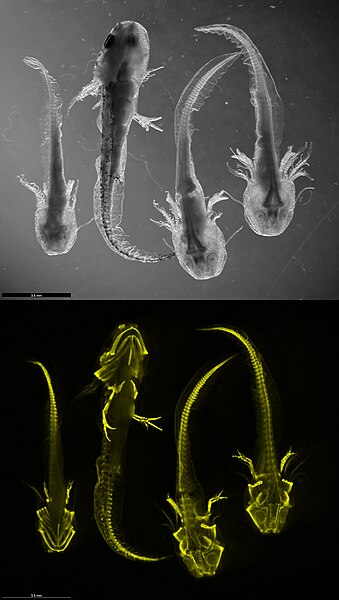
English: Top: Black and white bright-field microscopic view of four salamanders (Pleurodeles waltl) at developmental stages 35-37. Specimens 1 and 2 are captured ventrally, whereas specimens 3 and 4 are imaged dorsally. Among these, specimens 1, 3, and 4 are leucistic mutants, characterized by a knocked-out pigmentation gene, while specimen 2 is a wild-type, exhibiting no genetic mutations.
Bottom: The bottom panel illustrates the collagen expression patterns (CO2A1 gene) in these same salamanders, employing Hybridization Chain Reaction RNA Fluorescence In Situ Hybridization (HCR RNA-FISH) technology. The fluorescent visualization predominantly highlights the skeletal areas where collagen is abundantly present. Nonetheless, collagen expression is also observable in other regions, including the gills and on a distinct patch on top of the head. The image was captured by Lennart Rikk from the Regenerative Immunology Lab (https://leighnd.github.io/) using Leica M205 FCA microscope.Eesti: Üleval: Mustvalge valgusmikroskoobi pilt neljast salamandrist (Pleurodeles waltl) arengufaasides 35-37. Isendid 1 ja 2 on pildil ventraalselt, isendid 3 ja 4 dorsaalselt. Isendid 1, 3 ja 4 leukistilised mutandid, mida iseloomustab välja lülitatud pigmendigeen, samas kui isend 2 on metsiktüüpi, ilma geneetiliste mutatsioonideta.
All: Visualiseeritud on kollageeni ekspressioon (CO2A1 geen) samades salamandrites kasutades hübridisatsiooni ahelreaktsiooni RNA fluorestsentsi in situ hübridisatsiooni (HCR RNA-FISH) tehnoloogiat. Fluorestsents tõstab esile peamiselt luustiku piirkonna, kus kollageeni on rohkelt. Siiski on kollageeni ekspressioon märgatav ka teistes piirkondades nagu lõpustes ja pealeal eristuval laigul. Pildi tegi Lennart Rikk Regeneratiivse Immunoloogia Laborist (https://leighnd.github.io/) mikroskoobiga Leica M205 FCA. |
| Date | |
| Source | Own work |
| Author | Yodalr |
Licensing edit
- You are free:
- to share – to copy, distribute and transmit the work
- to remix – to adapt the work
- Under the following conditions:
- attribution – You must give appropriate credit, provide a link to the license, and indicate if changes were made. You may do so in any reasonable manner, but not in any way that suggests the licensor endorses you or your use.
| This file was uploaded as part of Wiki Science Competition 2023. |
File history
Click on a date/time to view the file as it appeared at that time.
| Date/Time | Thumbnail | Dimensions | User | Comment | |
|---|---|---|---|---|---|
| current | 21:58, 15 December 2023 | | 2,048 × 3,624 (3.06 MB) | Yodalr (talk | contribs) | Uploaded own work with UploadWizard |
You cannot overwrite this file.
File usage on Commons
The following 2 pages use this file:
File usage on other wikis
The following other wikis use this file:
- Usage on en.wikipedia.org
- Usage on et.wikipedia.org