File:Hippel Lindau.gif

Size of this preview: 508 × 599 pixels. Other resolutions: 204 × 240 pixels | 407 × 480 pixels | 805 × 949 pixels.
Original file (805 × 949 pixels, file size: 278 KB, MIME type: image/gif)
Captions
Captions
Add a one-line explanation of what this file represents
Summary
edit
| Description |
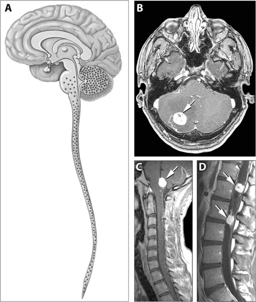
English: Distribution of Hemangioblastomas in the Central Nervous Systems of Study Patients
(A) Schematic representation of the distribution of CNS hemangioblastomas (red dots) in the 25 von Hippel-Lindau disease patients on MRI. Most (98%) of hemangioblastomas were found below the level of the tentorium in the cerebellum, brainstem, and spinal cord. (B–D) Contrast-enhanced MRI demonstrating representative locations of hemangioblastomas including the cerebellum (B), brainstem (C) and spinal cord (D). (B) Axial view through the cerebellum demonstrating a hyperintense enhancing hemangioblastoma (arrow) with surrounding edema (hypointense area surrounding the tumor) that frequently is associated with these lesions. (C) Sagittal view through the posterior fossa demonstrating a hyperintense enhancing brainstem (medullary) hemangioblastoma (arrow) with surrounding edema. (D) Sagittal view through the thoracic and lumbar spinal cord demonstrating two hyperintense enhancing hemangioblastomas (arrows). The superior tumor is associated with a large intraspinal cyst (syrinx) that is common with these neoplasms (arrowhead) |
| Source | http://medicine.plosjournals.org/perlserv/?request=get-document&doi=10.1371/journal.pmed.0040060 |
| Author |
Licensing
edit
|
This file is licensed under the Creative Commons Attribution 2.5 Generic license.
|
This file was published in a Public Library of Science journal. Their website states that the content of all PLOS journals is published under the Creative Commons Attribution 4.0 license (or its previous version depending on the publication date), unless indicated otherwise.
|
File history
Click on a date/time to view the file as it appeared at that time.
| Date/Time | Thumbnail | Dimensions | User | Comment | |
|---|---|---|---|---|---|
| current | 13:43, 31 May 2007 | | 805 × 949 (278 KB) | Filip em (talk | contribs) | Distribution of Hemangioblastomas in the Central Nervous Systems of Study Patients (A) Schematic representation of the distribution of CNS hemangioblastomas (red dots) in the 25 von Hippel-Lindau disease patients on MRI. Most (98%) of hemangioblastomas w |
You cannot overwrite this file.
File usage on Commons
There are no pages that use this file.
File usage on other wikis
The following other wikis use this file:
- Usage on ar.wikipedia.org
- Usage on bs.wikipedia.org
- Usage on ca.wikipedia.org
- Usage on de.wikipedia.org
- Usage on de.wikibooks.org
- Usage on el.wikipedia.org
- Usage on en.wikipedia.org
- Usage on hy.wikipedia.org
- Usage on it.wikipedia.org
- Usage on ja.wikipedia.org
- Usage on ru.wikipedia.org
- Usage on sk.wikipedia.org
- Usage on uz.wikipedia.org
- Usage on zh.wikipedia.org