File:Histopathology of Echinococcus granulosus hydatid cyst in a sheep 09G0016 lores.jpg

No higher resolution available.
Histopathology_of_Echinococcus_granulosus_hydatid_cyst_in_a_sheep_09G0016_lores.jpg (699 × 482 pixels, file size: 99 KB, MIME type: image/jpeg)
Captions
Captions
Add a one-line explanation of what this file represents
| Description |
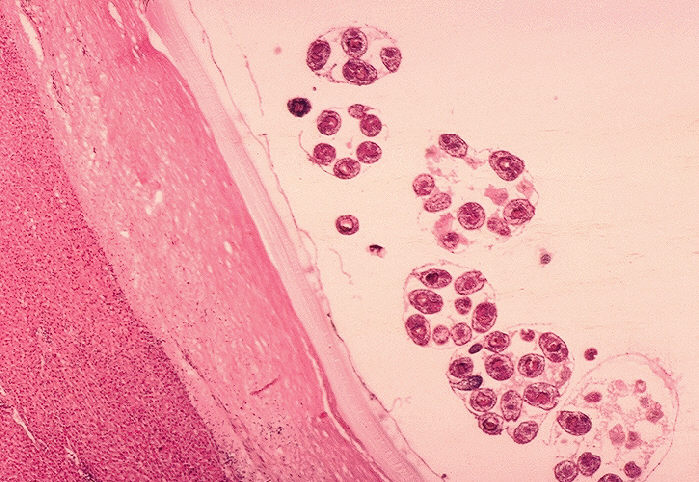
ID#: 910 Description: Histopathology of Echinococcus granulosus hydatid cyst in a sheep. Thick fibrous pericyst, hyaline ectocyst, and brood capsules filled with protoscolices are visible. Parasite. Content Providers(s): CDC/Dr.Peter Schantz Creation Date: 1975 Copyright Restrictions: None - This image is in the public domain and thus free of any copyright restrictions. As a matter of courtesy we request that the content provider be credited and notified in any public or private usage of this image. |
|||
| Source | http://phil.cdc.gov/PHIL_Images/03051999/00016/09G0016_lores.jpg | |||
| Author | ||||
| Permission (Reusing this file) |
|
File history
Click on a date/time to view the file as it appeared at that time.
| Date/Time | Thumbnail | Dimensions | User | Comment | |
|---|---|---|---|---|---|
| current | 14:01, 12 May 2006 | | 699 × 482 (99 KB) | Patho (talk | contribs) | {{Information| |Description=ID#: 910 Description: Histopathology of Echinococcus granulosus hydatid cyst in a sheep. Thick fibrous pericyst, hyaline ectocyst, and brood capsules filled with protoscolices are visible. Parasite. Content Providers(s): CDC/ |
You cannot overwrite this file.
File usage on Commons
The following page uses this file:
File usage on other wikis
The following other wikis use this file:
- Usage on de.wikibooks.org
- Usage on es.wikipedia.org
- Usage on fr.wikipedia.org
- Usage on hu.wikibooks.org
- Usage on la.wikipedia.org
- Usage on lt.wikipedia.org
- Usage on ru.wikipedia.org