File:Human brain left midsagitttal view closeup description 2-emphasizing-corpus-callosum.png
Human_brain_left_midsagitttal_view_closeup_description_2-emphasizing-corpus-callosum.png (702 × 491 pixels, file size: 1.32 MB, MIME type: image/png)
Captions
Captions
Summary edit
| Description |
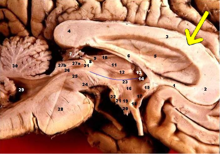
Human brain left - midsagitttal view - closeup. Emphasizing corpus callosum
On a half brain specimen, the Thalamus can be identified. The Thalamus (anteroom) is connected caudally with the midbrain and rostrally with the cerebral hemispheres. Note that the walls of the 3rd ventricle are completely formed by the Thalamus. The Hypothalamic Sulcus separates the Dorsal Thalamus superiorly from the Hypothalamus inferiorly. The subthalamus is located lateral to the hypothalamus and is not visible here. The anterior wall of the 3rd ventricle is a thin sheet of tissue called the Lamina Terminalis. In the Dorsal Thalamus note the Interthalamic Adhesion (Massa Intermedia), and the three parts of the Epithalamus: Stria Medullaris Thalami, Habenula, and the Pineal Gland. The floor of the Hypothalamus is made up of the Infundibulum, which connects with the pituitary gland, and posteriorly, the Tuber Cinereum and Mammillary Body.
|
| Date | |
| Source | http://www.healcentral.org/healapp/showMetadata?metadataId=40566 (Internet Archive of file description page) |
| Author |
John A Beal, PhD Dep't. of Cellular Biology & Anatomy, Louisiana State University Health Sciences Center Shreveport |
| Permission (Reusing this file) |
CC-BY |
| Other versions |
 |
Licensing edit
- You are free:
- to share – to copy, distribute and transmit the work
- to remix – to adapt the work
- Under the following conditions:
- attribution – You must give appropriate credit, provide a link to the license, and indicate if changes were made. You may do so in any reasonable manner, but not in any way that suggests the licensor endorses you or your use.
This file, which was originally posted to
http://www.healcentral.org/healapp/showMetadata?metadataId=40566, was reviewed on 25 September 2013 by reviewer Eleassar, who confirmed that it was available there under the stated license on that date.
|
File history
Click on a date/time to view the file as it appeared at that time.
| Date/Time | Thumbnail | Dimensions | User | Comment | |
|---|---|---|---|---|---|
| current | 20:30, 1 October 2009 | | 702 × 491 (1.32 MB) | Was a bee (talk | contribs) | {{Information| |Description='''Human brain left - midsagitttal view - closeup'''. Emphasizing corpus callosum # Corpus callosum, Rostrum # Corpus callosum, Genu # Corpus callosum, Corpus # Corpus callosum, Splenium # Septum pellucidum # Fornix, Corpus # |
You cannot overwrite this file.
File usage on Commons
There are no pages that use this file.
File usage on other wikis
The following other wikis use this file:
- Usage on ja.wikipedia.org