File:Intravital-Immunofluorescence-for-Visualizing-the-Microcirculatory-and-Immune-Microenvironments-in-pone.0057135.s009.ogv
Size of this JPG preview of this OGG file: 800 × 598 pixels. Other resolutions: 320 × 239 pixels | 640 × 478 pixels | 1,024 × 765 pixels | 1,280 × 956 pixels | 1,392 × 1,040 pixels.
Original file (Ogg Theora video file, length 9.6 s, 1,392 × 1,040 pixels, 2.13 Mbps, file size: 2.43 MB)
Captions
Captions
Add a one-line explanation of what this file represents
Summary edit
| Description |
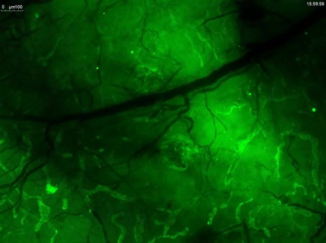
English: Lymphatic staining occurs within minutes after the application of secondary (donkey anti-rabbit Alexa 488, green) antibody on Lyve1-labeled tissue. Imaging started 2 minu after application of the secondary antibody. Duration: 18 min. |
||
| Date | |||
| Source | Video S6 from Kilarski W, Guc E, Teo J, Oliver S, Lund A, Swartz M (2013). "Intravital Immunofluorescence for Visualizing the Microcirculatory and Immune Microenvironments in the Mouse Ear Dermis". PLOS ONE. DOI:10.1371/journal.pone.0057135. PMID 23451163. PMC: 3581585. | ||
| Author | Kilarski W, Guc E, Teo J, Oliver S, Lund A, Swartz M | ||
| Permission (Reusing this file) |
|
||
| Provenance |
|
File history
Click on a date/time to view the file as it appeared at that time.
| Date/Time | Thumbnail | Dimensions | User | Comment | |
|---|---|---|---|---|---|
| current | 03:56, 8 March 2013 | 9.6 s, 1,392 × 1,040 (2.43 MB) | Open Access Media Importer Bot (talk | contribs) | Automatically uploaded media file from Open Access source. Please report problems or suggestions here. |
You cannot overwrite this file.
File usage on Commons
There are no pages that use this file.