File:Loupe-binoculaire-p1030891.jpg
Size of this preview: 800 × 594 pixels. Other resolutions: 320 × 238 pixels | 640 × 475 pixels | 1,024 × 760 pixels | 1,280 × 950 pixels | 2,535 × 1,882 pixels.
Original file (2,535 × 1,882 pixels, file size: 2.72 MB, MIME type: image/jpeg)
Captions
Captions
Add a one-line explanation of what this file represents
Summary
edit| Description |
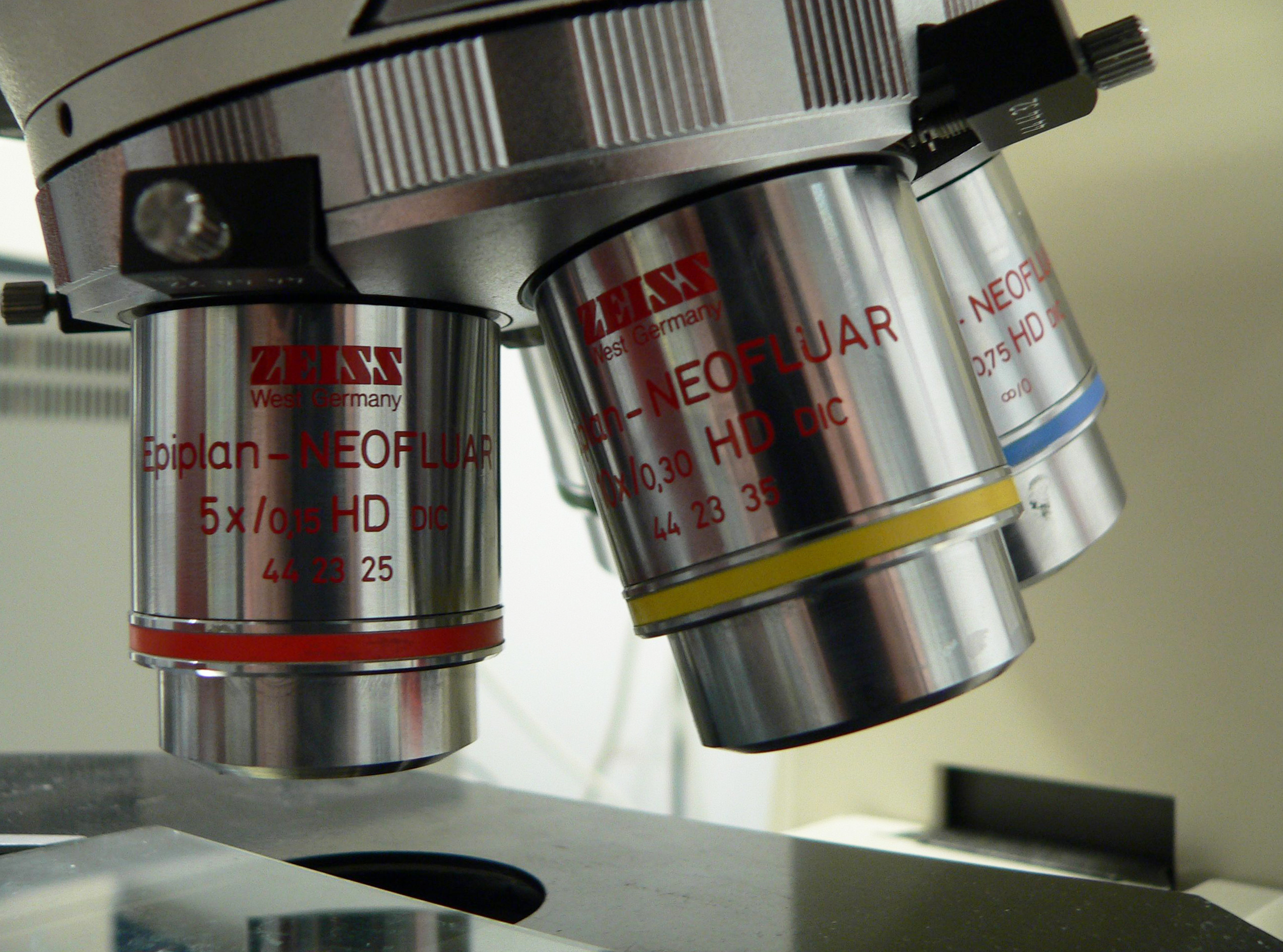
English: binocular microscope
Français : Loupe binoculaire |
||
| Date | Unknown date | ||
| Source | Own work | ||
| Author | Rama | ||
| Permission (Reusing this file) |
This file is licensed under the Creative Commons Attribution-Share Alike 2.0 France license.
 |

File history
Click on a date/time to view the file as it appeared at that time.
| Date/Time | Thumbnail | Dimensions | User | Comment | |
|---|---|---|---|---|---|
| current | 15:13, 19 December 2015 | | 2,535 × 1,882 (2.72 MB) | Jacek Halicki (talk | contribs) | tilt, more light, jpg compression |
| 13:43, 17 March 2006 |  | 2,560 × 1,920 (546 KB) | Rama (talk | contribs) | {{fr|Loupe binoculaire}} {{en|binocular microscope}} {{Rama}} Category:Microscopes |
You cannot overwrite this file.
File usage on Commons
The following 2 pages use this file:
File usage on other wikis
The following other wikis use this file:
- Usage on af.wikipedia.org
- Usage on ar.wikipedia.org
- Usage on bn.wikipedia.org
- Usage on en.wikipedia.org
- Microscopy
- Microscope
- Antonie van Leeuwenhoek
- Optical microscope
- Diffraction-limited system
- Objective (optics)
- Total internal reflection fluorescence microscope
- Fluorescence microscope
- Confocal microscopy
- Two-photon excitation microscopy
- Superlens
- 4Pi microscope
- Near-field scanning optical microscope
- STED microscopy
- Differential interference contrast microscopy
- Dark-field microscopy
- Bright-field microscopy
- Phase-contrast microscopy
- Köhler illumination
- Second-harmonic imaging microscopy
- Dispersion staining
- Vertico spatially modulated illumination
- Raman microscope
- Template:Optical microscopy
- Time-lapse microscopy
- Optical sectioning
- Sarfus
- Super-resolution microscopy
- Phase telescope
- Critical illumination
- Photoactivated localization microscopy
- Quantitative phase-contrast microscopy
- Light sheet fluorescence microscopy
- Live-cell imaging
- User:Egelberg/sandbox
- Lattice light-sheet microscopy
- American Microscopical Society
- User:Telementor/Userboxes/microscopy
- Talk:Microscope/Archive 1
- User:Lamals/sandbox/STED microscopy
- User:Lamals/sandbox/Fluorescence microscopy via coherent control
- Three-photon microscopy
- Usage on es.wikipedia.org
- Usage on fa.wikipedia.org
View more global usage of this file.