File:Micrograph of villous carcinoma, squamous carcinoma, clear cell carcinoma and hepatoid carcinoma.jpg

Size of this preview: 794 × 600 pixels. Other resolutions: 318 × 240 pixels | 636 × 480 pixels | 1,017 × 768 pixels | 1,280 × 967 pixels | 1,866 × 1,409 pixels.
Original file (1,866 × 1,409 pixels, file size: 1.06 MB, MIME type: image/jpeg)
Captions
Captions
Micrograph of villous carcinoma, squamous carcinoma, clear cell carcinoma and hepatoid carcinoma
Summary edit
| Description |
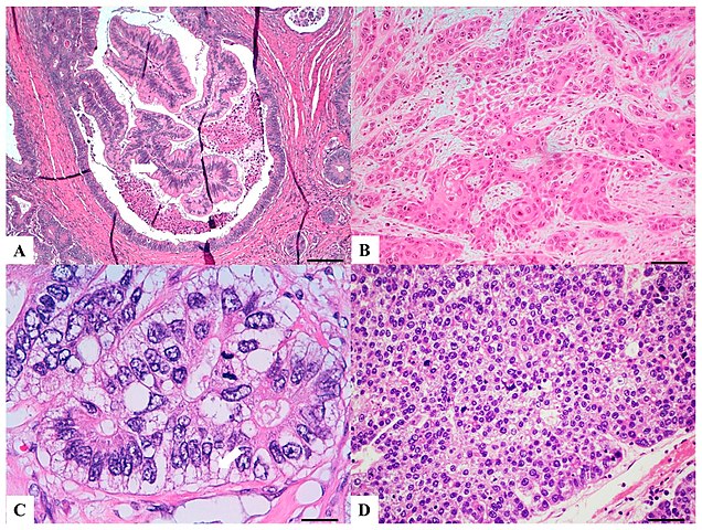
English: Original caption: "Haematoxylin and Eosin stained sections of rare type colorectal carcinomas. (A) Villous carcinoma: invasive carcinoma with villous features consisting of usually intraglandular papillary projections (yellow arrow) associated with an expansile growth pattern, at the deep portions of the tumor. Scale bar 400 micron. (B) Squamous carcinoma: morphologically similar to other squamous cell carcinomas occurring in other organs with possible keratinization. Scale bar 200 micron. (C) Clear cell carcinoma: clear cell cytoplasm identified in polygonal cells with a central nucleus, columnar cells with an eccentric nucleus (red arrow) and/or round/oval cells with abundant cytoplasm and inconspicuous marginally located nucleus similar to lipocytes or lipoblasts. Scale bar 50 micron. (D) Hepatoid carcinoma: large polygonal-shaped cells, with granular eosinophilic cytoplasm, prominent nucleoli and trabecular and pseudo-acinar growth pattern similar to hepatocarcinoma. Scale bar 200 micron." |
| Date | |
| Source |
(2019). "Morphology and Molecular Features of Rare Colorectal Carcinoma Histotypes". Cancers 11 (7): 1036. DOI:10.3390/cancers11071036. ISSN 2072-6694. |
| Author | Andrea Remo, Matteo Fassan, Alessandro Vanoli, Luca Reggiani Bonetti, Valeria Barresi, Fabiana Tatangelo, Roberta Gafà, Guido Giordano, Massimo Pancione, Federica Grillo and Luca Mastracci |
Licensing edit
This file is licensed under the Creative Commons Attribution 4.0 International license.
- You are free:
- to share – to copy, distribute and transmit the work
- to remix – to adapt the work
- Under the following conditions:
- attribution – You must give appropriate credit, provide a link to the license, and indicate if changes were made. You may do so in any reasonable manner, but not in any way that suggests the licensor endorses you or your use.
File history
Click on a date/time to view the file as it appeared at that time.
| Date/Time | Thumbnail | Dimensions | User | Comment | |
|---|---|---|---|---|---|
| current | 12:48, 30 September 2019 | | 1,866 × 1,409 (1.06 MB) | Mikael Häggström (talk | contribs) | User created page with UploadWizard |
You cannot overwrite this file.
File usage on Commons
The following 4 pages use this file:
- File:Micrograph of colorectal choriocarcinoma, rhabdoid colorectal carcinoma, carcinoma with osseous metaplasia and undifferentiated carcinoma.jpg
- File:Micrograph of lymphoepitelioma-like carcinoma, cribiform comedo-type carcinoma, micropapillary carcinoma and low grade tubulo-glandular carcinoma.jpg
- File:Micrograph of serrated adenocarcinoma, mucinous carcinoma, signet ring carcinoma and medullary carcinoma.jpg
- Template:Colorectal cancer histopathology types
File usage on other wikis
The following other wikis use this file:
- Usage on en.wikipedia.org