File:Neuropilo Corteza cerebro.png

Size of this preview: 774 × 599 pixels. Other resolutions: 310 × 240 pixels | 620 × 480 pixels | 992 × 768 pixels | 1,280 × 991 pixels | 2,172 × 1,682 pixels.
Original file (2,172 × 1,682 pixels, file size: 5.63 MB, MIME type: image/png)
Captions
Captions
Add a one-line explanation of what this file represents
Summary edit
| Description |
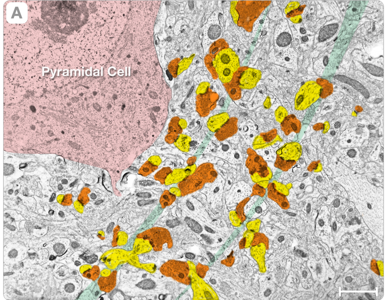
Español: Figura 1. El Neuropilo. Panel A es una vista de bajo aumento de un neuropilo en la corteza cerebral de rata. |
| Date | |
| Source | A Novel Synaptic Vesicle Fusion Path in the Rat Cerebral Cortex: The “Saddle” Point Hypothesis. (2014). PLoS ONE 9(6): e100710. doi:10.1371/journal.pone.0100710 |
| Author | Zampighi GA., Serrano R., Vergara JL. |
Licensing edit
This file is licensed under the Creative Commons Attribution 4.0 International license.
- You are free:
- to share – to copy, distribute and transmit the work
- to remix – to adapt the work
- Under the following conditions:
- attribution – You must give appropriate credit, provide a link to the license, and indicate if changes were made. You may do so in any reasonable manner, but not in any way that suggests the licensor endorses you or your use.
File history
Click on a date/time to view the file as it appeared at that time.
| Date/Time | Thumbnail | Dimensions | User | Comment | |
|---|---|---|---|---|---|
| current | 02:41, 14 July 2022 | | 2,172 × 1,682 (5.63 MB) | Sanador2.0 (talk | contribs) | Uploaded a work by Zampighi GA., Serrano R., Vergara JL. from A Novel Synaptic Vesicle Fusion Path in the Rat Cerebral Cortex: The “Saddle” Point Hypothesis. (2014). PLoS ONE 9(6): e100710. doi:10.1371/journal.pone.0100710 with UploadWizard |
You cannot overwrite this file.
File usage on Commons
There are no pages that use this file.
File usage on other wikis
The following other wikis use this file:
- Usage on es.wikipedia.org