File:Physiology and animal mechanism - first-book of natural history, prepared for the use of schools and colleges (1841) (14784391632).jpg
Original file(1,442 × 2,338 pixels, file size: 669 KB, MIME type: image/jpeg)

Captions

Captions

Add a one-line explanation of what this file represents

Summary edit

Description
English:

Identifier: 61350030R.nlm.nih.gov
Title: Physiology and animal mechanism : first-book of natural history, prepared for the use of schools and colleges
Year: 1841 (1840s)
Authors: Milne-Edwards, H. (Henri), 1800-1885 Comté, Achille, 1802-1866 Ruschenberger, W. S. W. (William Samuel Waithman), 1807-1895
Subjects: Physiological Phenomena
Publisher: Philadelphia : Turner & Fisher
Contributing Library: U.S. National Library of Medicine
Digitizing Sponsor: Open Knowledge Commons, U.S. National Library of Medicine

View Book Page: Book Viewer
About This Book: Catalog Entry
View All Images: All Images From Book
Click here to view book online to see this illustration in context in a browseable online version of this book.

Text Appearing Before Image:
punctalachrymalia.—c. n. the nasal canal. Fio. 6. The nasal fossse open and seen from the inside.—c. the craniums. f. the frontal sinuses, cavities hollowed out in the substance of the frontalbone, and in communication with the nasal fossse.—n. o. the olfactory nerver. its branches ramifying on the pituitary or schneiderian membrane.—-os,cm, ci, superior, middle and inferior turbinated bonos ; these are projectingplates of bone which serve to increase the extent of tho surface of the organof smell—ph. the swallow or pharynx—t, the opening of the Eustachian tube, Fig. 7. The larynx seen in front. The internal line indicates the shapeof the. internal surface of this organ.—»A. the hyoid bone.—I. the larynx, t, the trachea.—br. the bronchioe.—c. v. vocal cords. Fig. 8. The larynx open, seen from one side.—e. the epiglottis.—v. oneof the ventricles of the larynx.—Ig. the superior ligament of the larynx.—.c, v. one of the vocal cords,—U the trachea, Plate 6.
Text Appearing After Image:
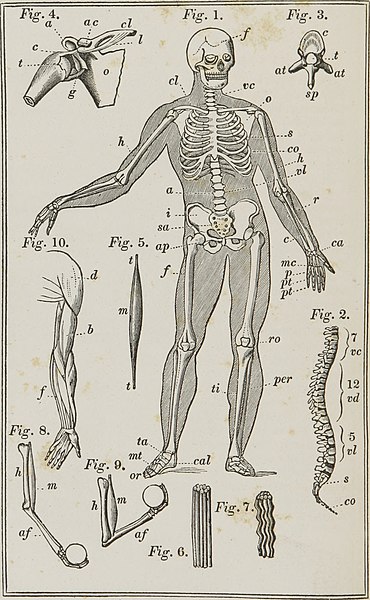
Organs of Motion. EXPLANATION OF PLATE 6. Fig. 1. Skeleton of man; the external line indicates the shape of thebody—/. the frontal bone—v. c. the cervical vertebrae—s. the sternum orbreast bone—co. the ribs—v. I. the lumbar vertebrae—a. the abdomen—sa.the sacrum—cl. the clavicle—o. the scapula or shoulder blade—h. the hu-merus—r. the radius—c. the cubitus or ulna—ca. the carpus—mc. the me-tacarpus—p. and pt. the phalanges or bones of the fingers—i. the ilium—/. the femur-—ro. the rotula, patella or knee-pan—ti. the tibia—per. the fibula—ta. the tarsus—mt. the metatarsus—or. the toes—cal. the calcis or heel. Fig. 2. The vertebral column seen in profile—vc. the cervical regioncomposed of seven vertebrae—v. d. the dorsal region—vl. the lumbar region—s. the sacrum—co. coxyx. Fig. 3. A vertebra seen from above—c. body of the vertebra—t. a holefor the passage of the spinal marrow—a. t. transverse processes— s. p. spi-nous pro

Note About Images

Please note that these images are extracted from scanned page images that may have been digitally enhanced for readability - coloration and appearance of these illustrations may not perfectly resemble the original work.
Date
Source

https://www.flickr.com/photos/internetarchivebookimages/14784391632/

Author Internet Archive Book Images
Permission
(Reusing this file)
At the time of upload, the image license was automatically confirmed using the Flickr API. For more information see Flickr API detail.
Flickr tags
InfoField
Flickr posted date
InfoField
30 July 2014


Licensing edit

This image was taken from Flickr's The Commons. The uploading organization may have various reasons for determining that no known copyright restrictions exist, such as:
  1. The copyright is in the public domain because it has expired;
  2. The copyright was injected into the public domain for other reasons, such as failure to adhere to required formalities or conditions;
  3. The institution owns the copyright but is not interested in exercising control; or
  4. The institution has legal rights sufficient to authorize others to use the work without restrictions.

More information can be found at https://flickr.com/commons/usage/.


Please add additional copyright tags to this image if more specific information about copyright status can be determined. See Commons:Licensing for more information.
This image was originally posted to Flickr by Internet Archive Book Images at https://flickr.com/photos/126377022@N07/14784391632. It was reviewed on 26 October 2015 by FlickreviewR and was confirmed to be licensed under the terms of the No known copyright restrictions.

26 October 2015

File history

Click on a date/time to view the file as it appeared at that time.

Date/TimeThumbnailDimensionsUserComment
current23:45, 26 October 2015Thumbnail for version as of 23:45, 26 October 20151,442 × 2,338 (669 KB) (talk | contribs)== {{int:filedesc}} == {{information |description={{en|1=<br> '''Identifier''': 61350030R.nlm.nih.gov<br> '''Title''': [https://www.flickr.com/photos/internetarchivebookimages/tags/bookid61350030R.nlm.nih.gov Physiology and animal mechanism : first-boo...

There are no pages that use this file.