File:Rabies encephalitis Negri bodies PHIL 3377 lores.jpg

Size of this preview: 800 × 532 pixels. Other resolutions: 320 × 213 pixels | 640 × 425 pixels | 1,024 × 681 pixels | 1,280 × 851 pixels | 1,801 × 1,197 pixels.
Original file (1,801 × 1,197 pixels, file size: 438 KB, MIME type: image/jpeg)
Captions
Captions
Add a one-line explanation of what this file represents
Summary edit
| Description |
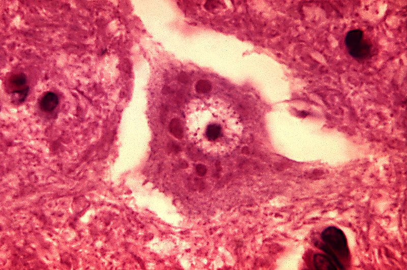
English: This micrograph depicts the histopathologic changes associated with rabies encephalitis prepared using an H&E stain.
Note the Negri bodies, which are cellular inclusions found most frequently in the pyramidal cells of Ammon's horn, and the Purkinje cells of the cerebellum. They are also found in the cells of the medulla and various other ganglia. |
||
| Date | |||
| Source |
|
||
| Author | Content Provider(s): CDC/Dr. Daniel P. Perl | ||
| Permission (Reusing this file) |
Copyright Restrictions: None – This image is in the public domain and thus free of any copyright restrictions. As a matter of courtesy we request that the content provider be credited and notified in any public or private usage of this image. | ||
| Other versions |
 |
Licensing edit
This image is a work of the Centers for Disease Control and Prevention, part of the United States Department of Health and Human Services, taken or made as part of an employee's official duties. As a work of the U.S. federal government, the image is in the public domain.
|
File history
Click on a date/time to view the file as it appeared at that time.
| Date/Time | Thumbnail | Dimensions | User | Comment | |
|---|---|---|---|---|---|
| current | 07:27, 26 October 2011 | | 1,801 × 1,197 (438 KB) | Ghainmem (talk | contribs) | Higher-resolution version |
| 18:01, 30 May 2006 |  | 700 × 465 (57 KB) | Patho (talk | contribs) | {{Information| |Description=ID#: 3377 Description: This micrograph depicts the histopathologic changes associated with rabies encephalitis prepared using an H&E stain. Note the Negri bodies, which are cellular inclusions found most frequently in the pyra |
You cannot overwrite this file.
File usage on Commons
The following page uses this file:
File usage on other wikis
The following other wikis use this file:
- Usage on ar.wikipedia.org
- Usage on de.wikibooks.org
- Usage on fr.wikipedia.org
- Usage on ja.wikipedia.org
- Usage on th.wikipedia.org
- Usage on uk.wikipedia.org