Thu Jun 18 16:24:37 CEST 2015 edit
errors edit
- File:ND25.jpg aborted: duplicate: File:Nita_Wallenberg_1917.jpg
- File:ND34_Midsommarvaka.jpg aborted: duplicate: File:Nils_Dardel_Midsommarvaka.jpg
- File:ND47Bergman.jpg aborted: duplicate: File:Nils_Dardel_Hjalmar_Bergman.jpg
- File:ND51Gaby.jpg aborted: duplicate: File:Nils_Dardel_Gaby.jpg
- File:ND52TollieZellman.jpg aborted: duplicate: File:Tollie_Zellman_målad_av_Nils_Dardel.jpg
- File:ND55.jpg aborted: duplicate: File:Nils_Dardel_Eva_Bonnier.jpg
- File:ND91.jpg aborted: duplicatepain: File:Nils_Dardel_Paranoikern.jpg
- File:ND97.jpg aborted: duplicate: File:Nils_Dardel_John_Blund.jpg
- File:ND_Nybroviken.jpg aborted: duplicate: File:Nils_Dardel_Nybroviken.jpg
- File:Thora.jpg aborted: duplicate: File:Nils_Dardel_Thora.jpg


Thu Jun 18 14:25:07 CEST 2015 edit
errors edit
- File:Grant_1962_192.png exists-normalized
- File:Grant_1962_193.png exists-normalized
- File:Grant_1962_196.png exists-normalized
- File:Grant_1962_199.png exists-normalized
- File:Grant_1962_208.png exists-normalized
- File:Grant_1962_223.png exists-normalized
- File:Grant_1962_230.png exists-normalized
- File:Grant_1962_237.png exists-normalized


Thu Jun 18 13:42:52 CEST 2015 edit
errors edit
- File:Grant_1962_236.png aborted: exists: File:Grant_1962_236.png

Thu Jun 18 11:50:55 CEST 2015 edit
errors edit
- File:Grant_1962_181.png exists-normalized
- File:Grant_1962_183.png exists-normalized
- File:Grant_1962_185.png exists-normalized
- File:Grant_1962_188.png exists-normalized
- File:Grant_1962_191.png exists-normalized


Thu Jun 18 09:58:19 CEST 2015 edit
errors edit
- File:Grant_1962_163.png exists-normalized


Wed Jun 17 11:17:26 CEST 2015 edit
errors edit
- File:Grant_1962_136.png exists-normalized
- File:Grant_1962_137.png exists-normalized
- File:Grant_1962_146.png exists-normalized


Fri Jun 12 13:19:02 CEST 2015 edit
-
 Artwork Details Dimensions: 9.62 X 12.25 in (24.43 X 31.12 cm) Medium: oil on canvas Creation Date: 18th Century German School, 18th Century Hippolytus, Phaedra and Theseus

Fri Jun 12 13:17:58 CEST 2015 edit

Fri Jun 12 13:16:42 CEST 2015 edit
errors edit
- File:Salomon_Koninck_A_SCHOLAR_IN_HIS_STUDY badfilename
- File:Samuel_van_Hoogstraten_The_Visitation.jpeg aborted: exists: File:Samuel_van_Hoogstraten_The_Visitation.jpeg
-
![Samuel van Hoogstraten The Visitation Dimensions: 230 by 178 mm Medium: Pen and brown ink with brown and red wash within brown ink framing lines "Provenance Schneider Collection, 1876 (according to Sumowski); M.A. Marmontel, his sale, Paris, 25-26 January 1883, lot 245 (as Rembrandt); sale, Paris, 28-29 March 1898, lot 54; Professor E. Perman, Stockholm, thence by descent to the present owner[1]"](https://upload.wikimedia.org/wikipedia/commons/thumb/5/5b/Samuel_van_Hoogstraten_The_Visitation.jpeg/95px-Samuel_van_Hoogstraten_The_Visitation.jpeg) Samuel van Hoogstraten The Visitation Dimensions: 230 by 178 mm Medium: Pen and brown ink with brown and red wash within brown ink framing lines "Provenance Schneider Collection, 1876 (according to Sumowski); M.A. Marmontel, his sale, Paris, 25-26 January 1883, lot 245 (as Rembrandt); sale, Paris, 28-29 March 1898, lot 54; Professor E. Perman, Stockholm, thence by descent to the present owner[1]"
Samuel van Hoogstraten The Visitation Dimensions: 230 by 178 mm Medium: Pen and brown ink with brown and red wash within brown ink framing lines "Provenance Schneider Collection, 1876 (according to Sumowski); M.A. Marmontel, his sale, Paris, 25-26 January 1883, lot 245 (as Rembrandt); sale, Paris, 28-29 March 1898, lot 54; Professor E. Perman, Stockholm, thence by descent to the present owner[1]"
![Samuel van Hoogstraten The Visitation Dimensions: 230 by 178 mm Medium: Pen and brown ink with brown and red wash within brown ink framing lines "Provenance Schneider Collection, 1876 (according to Sumowski); M.A. Marmontel, his sale, Paris, 25-26 January 1883, lot 245 (as Rembrandt); sale, Paris, 28-29 March 1898, lot 54; Professor E. Perman, Stockholm, thence by descent to the present owner[1]"](/wiki/File:Samuel_van_Hoogstraten_The_Visitation.jpeg)
Fri Jun 12 13:15:05 CEST 2015 edit
-
Samuel van Hoogstraten The Visitation Dimensions: 230 by 178 mm Medium: Pen and brown ink with brown and red wash within brown ink framing lines "Provenance Schneider Collection, 1876 (according to Sumowski); M.A. Marmontel, his sale, Paris, 25-26 January 1883, lot 245 (as Rembrandt); sale, Paris, 28-29 March 1898, lot 54; Professor E. Perman, Stockholm, thence by descent to the present owner[2]"
Fri Jun 12 13:11:39 CEST 2015 edit
-
 Juan Bautista Martinez del Mazo Portrait of Diego de Silva y Velazquez Artwork Details Dimensions: 47.75 X 38.5 in (121.28 X 97.79 cm) Medium: oil on canvas

Fri Jun 12 13:10:42 CEST 2015 edit
errors edit
-
Juan Bautista Martinez del Mazo Portrait of Diego de Silva y Velazquez Artwork Details Dimensions: 47.75 X 38.5 in (121.28 X 97.79 cm) Medium: oil on canvas
Tue Jul 15 22:06:28 CEST 2014 edit
-
 LABIAL MUCOSA: (Figures 3 and 4) With the patient's mouth partially open, visually examine the labial mucosa and sulcus of the maxillary vestibule and frenum and the mandibular vestibule. Observe the color, texture, and any swelling or other abnormalities of the vestibular mucosa and gingiva.
 -
 FACE: (Figure 1) The extraoral assessment includes inspection of the face, head, and neck. The face, ears, and neck are observed, noting any asymmetry or changes on the skin such as crusts, fissuring, growths, and/or color change. The regional lymph node areas are bilaterally palpated to detect any enlarged nodes, and if detected, their mobility and consistency. A recommended order of examination includes the preauricular, submandibular, anterior cervical, posterior auricular, and posterior cervical regions.
 -
 (Figure 10) Grasping the tip of the tongue with a piece of gauze will assist full protrusion and will aid examination of the more posterior aspects of the tongue's lateral borders
 -
 (Figure 11) Then examine the ventral surface. Palpate the tongue to detect growths.
 -
 FLOOR: (Figure 12) With the tongue still elevated, inspect the floor of the mouth for changes in color, texture, swellings, or other surface abnormalities.
 -
 PALATE: (Figures 13 and 14) With the mouth wide open and the patient's head tilted back, gently depress the base of the tongue with a mouth mirror. First inspect the hard and then the soft palate.
 -
 (Figure 14) Examine all soft palate and oropharyngeal tissues.
 -
 (Figure 15) Bimanually palpate the floor of the mouth for any abnormalities. All mucosal or facial tissues that seem to be abnormal should be palpated.
 -
 Homogenous leukoplakia in the floor of the mouth in a smoker. Biopsy showed hyperkeratosis.
 -
 Clinically, a leukoplakia on left buccal mucosa. However, the biopsy showed early squamous cell carcinoma. The lesion is suspicious because of the presence of nodules.
 -
 Nodular leukoplakia in right commissure. Biopsy showed severe epithelial dysplasia.
 -
 Erythroleukoplakia in left commissure and buccal mucosa. Biopsy showed mild epithelial dysplasia and presence of candida infection. A 2-3 week course of anti-fungal treatment may turn this type of lesion into a homogenous leukoplakia.
 -
 LIPS: (Figure 2) Begin examination by observing the lips with the patient's mouth both closed and open. Note the color, texture and any surface abnormalities of the upper and lower vermilion borders.
 -
 LABIAL MUCOSA: (Figures 3 and 4) With the patient's mouth partially open, visually examine the labial mucosa and sulcus of the maxillary vestibule and frenum and the mandibular vestibule. Observe the color, texture, and any swelling or other abnormalities of the vestibular mucosa and gingiva.
 -
 BUCCAL MUCOSA: (Figures 5 and 6) Retract the buccal mucosa. Examine first the right then the left buccal mucosa extending from the labial commissure and back to the anterior tonsillar pillar. Note any change in pigmentation, color, texture, mobility, and other abnormalities of the mucosa, making sure that the commissures are examined carefully and are not covered by the retractors during the retraction of the cheek.
 -
 GINGIVA: (Figure 7) First, examine the buccal and labial aspects of the gingiva and alveolar ridges (processes) by starting with the right maxillary posterior gingiva and alveolar ridge and then move around the arch to the left posterior area. Drop to the left mandibular posterior gingiva and alveolar ridge and move around the arch to the right posterior area.
 -
 TONGUE: (Figure 8) With the patient's tongue at rest, and mouth partially open, inspect the dorsum of the tongue for any swelling, ulceration, coating, or variation in size, color, or texture. Also note any change in the pattern of the papillae covering the surface of the tongue and examine the tip of the tongue. The patient should then protrude the tongue, and the examiner should note any abnormality of mobility or positioning.
 -
 (Figure 9) With the aid of mouth mirrors, inspect the right and left lateral margins of the tongue.


Tue Jul 08 15:45:55 CEST 2014 edit


Mon Jul 07 13:58:37 CEST 2014 edit

Mon Jul 07 09:01:38 CEST 2014 edit
errors edit
- File:Gluteus_maximus.gif exists-normalized


Mon Jul 07 08:59:17 CEST 2014 edit
errors edit
- File:Gluteus_maximus.gif exists-normalized
- File:Pectineus.gif exists-normalized
- File:Quadriceps.gif exists-normalized
- File:Rectus_femoris.gif exists-normalized
- File:Sartorius.gif exists-normalized
Mon Jul 07 08:57:45 CEST 2014 edit
errors edit
- File:Gluteus_maximus.gif exists-normalized
- File:Pectineus.gif exists-normalized
- File:Quadriceps.gif exists-normalized
- File:Rectus_femoris.gif exists-normalized
- File:Sartorius.gif exists-normalized
Mon Jul 07 08:56:07 CEST 2014 edit
errors edit
- File:Gluteus_maximus.gif exists-normalized
- File:Pectineus.gif exists-normalized
- File:Quadriceps.gif exists-normalized
- File:Rectus_femoris.gif exists-normalized
- File:Sartorius.gif exists-normalized


Fri Mar 21 11:01:11 CET 2014 edit
errors edit
- File:Grant_1962_71.png exists-normalized
- File:Grant_1962_86.png exists-normalized
Fri Mar 21 10:26:12 CET 2014 edit
errors edit
- File:Grant_1962_71.png exists-normalized
- File:Grant_1962_86.png exists-normalized


Thu Mar 13 07:44:12 CET 2014 edit


Tue Feb 04 11:48:13 CET 2014 edit
errors edit
- File:Lawrence_1960_5.13_F.png aborted: exists: File:Lawrence_1960_5.13_F.png


Tue Feb 04 10:04:19 CET 2014 edit
errors edit
- File:Lawrence_1960_25.png exists-normalized


Tue Feb 04 09:32:34 CET 2014 edit

Mon Jan 27 22:25:49 CET 2014 edit
errors edit
- File:Lawrence_1960_19.13.png aborted: exists: File:Lawrence_1960_19.13.png


Mon Jan 27 11:39:59 CET 2014 edit
-
 Evolution of the neuron
 -
 Evolution of the central nervous system
 -
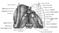
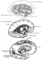
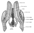
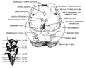
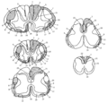
 A. Dorsal view of the brain of the dogfish. S. Dorsal view of the brain of the dogfish with the ventricles open. (Adapted from Ronton and Clark, The Anafomy of fhe Nervous System, 10th ed., W. B. Saunders Company, Philadelphia, 1959.)
 -
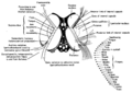
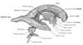
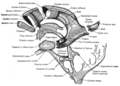
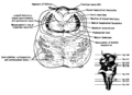
 Above, lateral view of the brain of the dogfish. Be/ow, midsagittal section of the brain of the dogfish. (Adapted from Ranson and Clark, The Anatomy of the Nervous System, 10th ed., W. B. Saunders Company, Philadelphia, 1959.)
 -
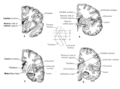
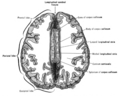
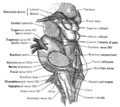
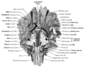
 Lateral view of the human cerebral hemispheres illustrating the principal gyri.
 -

-

-

-

-

-
-
-
-
-

-
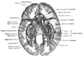
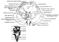
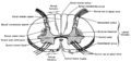
 Lateral view of the human cerebral hemispheres illustrating the principal sulci and lobes.
 -

-
-
-

-

-

-

-
-

-

-

-
-
-

-
-

-

-

-
-

-

-
-
-

-

-

-

-
-

-

-

-

-

-

-

-

-

-

-
-

-

-
-

-

-
-

-


Sat Dec 14 11:40:20 CET 2013 edit
errors edit
- File:Phineas_gage_-_1868_skull_diagram.jpg aborted: exists: File:Phineas_gage_-_1868_skull_diagram.jpg


_Nodule.jpg)


.jpg)


_Is_Broken_Down_Into_Monoglycerides_(b).jpg)


2015-08-17 02:57:34 edit
-
 == Summary ==
Paul Signac: Women at the Well Artist Paul Signac (1863–1935) 


Alternative names Hsi-nieh-kʻo; Polʹ Sinʹi︠a︡k; Paul Victor Jules Signac; Signac; p. signac; signac p.Description French painter, drawer, aquarellist and printmaker Date of birth/death 11 November 1863 
15 August 1935 Location of birth/death Paris Paris Work period from 1882 until 1935 Work location Authority file - : Q151573
- VIAF: 46767282
- ISNI: 0000000121309566
- ULAN: 500008410
- LCCN: n80005086
- NLA: 35499010
- WGA: s/signac
- Open Library: OL797509A
- GND: 119425157
- SELIBR: 213145
- SUDOC: 02713783X
- BNF: 119247303
- NDL: 00456562
- BIBSYS: 90127034
- NKC: ola2002151596
- SBN: MODV077499
- BNE: XX849634
- CiNii: DA05142749
- KulturNav: 8bf0923c-139c-48d6-96c9-f53c0d6bb064
- RKD: 72519
- J9U: 987007268225805171
- Koninklijke: 071281126
- Enciclopédia Itaú Cultural: pessoa351542/paul-signac
- WorldCat
Title Women at the WellDate 1892 Medium oil on canvas Dimensions 195 × 131 cm (76.7 × 51.5 in) Collection Musée d'Orsay Notes Contrary to all other versions on the web, the Metropolitan Museum of Art website has a mirror-inverted image of that painting: http://www.metmuseum.org/special/Signac/63.L.htm Please notifiy the uploader at User talk:AndreasPraefcke if you are sure which direction is the right one. Source/Photographer Image and original data provided by Erich Lessing Culture and Fine Arts Archives/ART RESOURCE, N.Y.

2015-08-18 15:42:27 edit
-
 == Summary ==
Paul Signac: Women at the Well Artist Paul Signac (1863–1935) Alternative names Hsi-nieh-kʻo; Polʹ Sinʹi︠a︡k; Paul Victor Jules Signac; Signac; p. signac; signac p.Description French painter, drawer, aquarellist and printmaker Date of birth/death 11 November 1863 15 August 1935 Location of birth/death Paris Paris Work period from 1882 until 1935 Work location Authority file - : Q151573
- VIAF: 46767282
- ISNI: 0000000121309566
- ULAN: 500008410
- LCCN: n80005086
- NLA: 35499010
- WGA: s/signac
- Open Library: OL797509A
- GND: 119425157
- SELIBR: 213145
- SUDOC: 02713783X
- BNF: 119247303
- NDL: 00456562
- BIBSYS: 90127034
- NKC: ola2002151596
- SBN: MODV077499
- BNE: XX849634
- CiNii: DA05142749
- KulturNav: 8bf0923c-139c-48d6-96c9-f53c0d6bb064
- RKD: 72519
- J9U: 987007268225805171
- Koninklijke: 071281126
- Enciclopédia Itaú Cultural: pessoa351542/paul-signac
- WorldCat
Title Women at the WellDate 1892 Medium oil on canvas Dimensions 195 × 131 cm (76.7 × 51.5 in) Collection Musée d'Orsay Notes Contrary to all other versions on the web, the Metropolitan Museum of Art website has a mirror-inverted image of that painting: http://www.metmuseum.org/special/Signac/63.L.htm Please notifiy the uploader at User talk:AndreasPraefcke if you are sure which direction is the right one. Source/Photographer Image and original data provided by Erich Lessing Culture and Fine Arts Archives/ART RESOURCE, N.Y.

2015-08-18 15:43:53 edit
-
 == Summary ==
Paul Signac: Women at the Well Artist Paul Signac (1863–1935) Alternative names Hsi-nieh-kʻo; Polʹ Sinʹi︠a︡k; Paul Victor Jules Signac; Signac; p. signac; signac p.Description French painter, drawer, aquarellist and printmaker Date of birth/death 11 November 1863 15 August 1935 Location of birth/death Paris Paris Work period from 1882 until 1935 Work location Authority file - : Q151573
- VIAF: 46767282
- ISNI: 0000000121309566
- ULAN: 500008410
- LCCN: n80005086
- NLA: 35499010
- WGA: s/signac
- Open Library: OL797509A
- GND: 119425157
- SELIBR: 213145
- SUDOC: 02713783X
- BNF: 119247303
- NDL: 00456562
- BIBSYS: 90127034
- NKC: ola2002151596
- SBN: MODV077499
- BNE: XX849634
- CiNii: DA05142749
- KulturNav: 8bf0923c-139c-48d6-96c9-f53c0d6bb064
- RKD: 72519
- J9U: 987007268225805171
- Koninklijke: 071281126
- Enciclopédia Itaú Cultural: pessoa351542/paul-signac
- WorldCat
Title Women at the WellDate 1892 Medium oil on canvas Dimensions 195 × 131 cm (76.7 × 51.5 in) Collection Musée d'Orsay Notes Contrary to all other versions on the web, the Metropolitan Museum of Art website has a mirror-inverted image of that painting: http://www.metmuseum.org/special/Signac/63.L.htm Please notifiy the uploader at User talk:AndreasPraefcke if you are sure which direction is the right one. Source/Photographer Image and original data provided by Erich Lessing Culture and Fine Arts Archives/ART RESOURCE, N.Y.
