User:Dcrjsr/gallery of protein structure
The first section is a collection of the original ribbon diagrams hand-drawn around 1980 by Jane Shelby Richardson, some hand-colored by her. Most of them were photographed by David C. Richardson for reproduction in "The Anatomy and Taxonomy of Protein Structures" Advances in Protein Chemistry 34:167-339, and most were later scanned and cleaned up by Claudia J. Richardson for the online version of "Anatax" at http://kinemage.biochem.duke.edu/teaching/anatax
The second section is later computer-graphics images by User:Dcrjsr, mostly made in the kinemage-display programs Mage (by David Richardson) or KiNG (by Ian Davis and Vincent Chen).
- Protein Ribbon Drawings by User:Dcrjsr
-
 Staph nuclease B&W shaded ribbon
 -
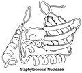 Staphylococcal nuclease ribbon, from 1sns
 -
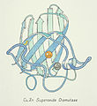 Cu,Zn superoxide dismutase, 2sod
 -
 Cu,Zn SOD ribbon drawing, white background
 -
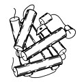
 Myohemerythrin ribbon 4-helix bundle, from 2mhr
 -
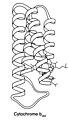 Cytochrome b562 ribbon 4-helix bundle, from 156b
 -
 Sketch drawing of myoglobin
 -
 Hemoglobin beta-subunit ribbon, red
 -
 Hemoglobin beta subunit ribbon, white background
 -
 Hen-egg-white lysozyme, ribbon schematic
 -
 Photosynthetic Reaction Center drawing, 1prc
 -
 TIM ribbon pastel matted, PDB file 1tim
 -
 TIM ribbon pastel, wide mat
 -
 Ribbon drawing of triose phosphate isomerase (TIM)
 -
 TIM ribbon pastel, white background
 -
 TIM ribbon B&W line, end view, 1tim
 -
 TIM ribbon B&W shaded, end view, 1tim
 -
 LDH domain 1 "Rossmann fold" ribbon, from 2ldh
 -
 LDH domain 1, prototype Rossmann fold, from 2ldh
 -
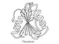 Flavodoxin ribbon B&W line, 1fxn
 -
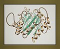
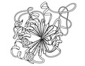
 Flavodoxin worm drawing, with flavin, from 1fxn
 -
 Glycogen phosphorylase, domain 2, 5-layered
 -
 Carboxypeptidase A (PDB 1cpa) ribbon schematic
 -
 Subtilisin BPN', ribbon diagram (PDB 1sbt)
 -
 PHBH, 3 domains
 -
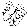
 Immunoglobulin VL ribbon, from 1rei
 -
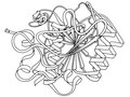
 RibonucleaseS ribbon drawing scratchboard
 -
 RibonucleaseA SS gold, line, 7rsa
 -
 RibonucleaseA SS pale ribbon, 7rsa
 -
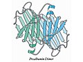 Prealbumin dimer ribbon, 2pab
 -
 Catabolite gene activator protein dimer
 -
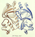
 CAP protein Greek-key paired beta strands
 -
 Elastase (Ser protease) ribbon, from 1est
 -
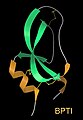
 Trypsin inhibitor (BPTI) ribbon schematic, SS yellow
 -
 BPTI ribbon diagram, hand-drawn from 2pti
 -
 Potato carboxypeptidase inhibitor, from 4cpa
 -
 Insulin monomer "worm" drawing, from 1ins
 -
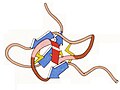
 Cytochrome C (PDB 1cyt) ribbon schematic
 -
 Rubredoxin (PDB 2rxn) ribbon diagram
 -
 Two common protein "folds": TIM & LDH
 -
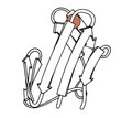
 Drawing a twisted-arrow ribbon
 -
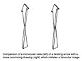
 Offset-lines illusion for beta ribbons
 -
 Hand-drawn helix ribbons at various angles
 -
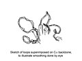 Loop smoothing for ribbon diagrams
 -
 Ribbon-drawing technique, two examples
 -
 Composite of ribbon-drawing representations: helix, sheet & loops
 -
 Betabellin design model, beta sandwich
 -
 Felix design model, 4-helix bundle


- Protein-Fold Computer Graphics Images by User:Dcrjsr
-
 Early multi-strand computer ribbons (vector) - 1wrp
 -
 Staphylococcal nuclease with Ca and PdtP, 3d6m
 -
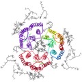
 Yeast Cu,Zn SOD dimer ribbon, 1sdy
 -
 Cu,Zn superoxide dismutase, crystal au
 -
 Human Cu,Zn SOD1 rib &site PDB 2c9v
 -
 Domain1 of human mitochondrial MnSOD 1n0j
 -
 Domain2 of human mitochondrial MnSOD 1n0j
 -
 Ribonuclease A ribbon, 7rsa
 -
 RNase S superimposed on RNase A
 -
 RNAse-inhibitor protein binding RNAse, 1dfj
 -
 HIV-1 protease dimer, from 3phv
 -
 GFP ribbon & fluorophore, 1ema
 -
 Bacteriorhodopsin trimer: ribbon, retinol, lipids (2brd)
 -
 Alpha-hemolysin 7-mer, down pore, 7ahl
 -
 Alpha-hemolysin 7-mer, side view, 7ahl
 -
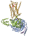
 b2-adrenergic receptor structure, from 2rh1
 -
 beta2-adrenergic receptor with its G protein, 3sn6
 -
 Photosystem II at FEL, from 4fby
 -
 Kinesin heads & neck ribbons, 3kin
 -
 Tubulin dimer, from 1tub
 -
 Ankyrin, 12 2-helix repeats (from 1n11)
 -
 BPTI sequence and folded structure
 -
 BPTI ribbon with SS, from 1bpi
 -
 BPTI swish ribbon, 1bpi
 -
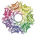
 Righthanded 3-side beta helix, pectate lyase C
 -
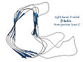
 Lefthanded 3-side beta helix from 1qre
 -
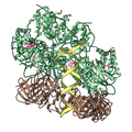
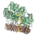
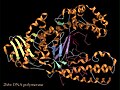 DNA polymerase complex, 2hhv
 -
 HLA antigen of the MHC complex, 3HLA
 -
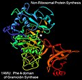 1amu gramicidin synthase PheA rib
 -
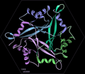
 3dcx, pentamer with tighter between-chain contacts
 -
 Trimer ribbon, 1xk8 from SECSG
 -
 Icosahedral virus capsid trimer of SBMV
 -
 Vault cell compartment half, side view, 2zuo,2zv4,2zv5
 -
 Vault cell compartment half, end view, 2zuo,2zv4,2zv5
 -
 Clamp-loader/DNA/clamp: open lock-washer conf, 3u5z
 -
 Clamp-loader/DNA/sliding clamp: closed ring conf, 3u5z
 -
 structure of 9-subunit human exosome, from 2nn6


