User:IsadoraofIbiza/Portfolio

These are several planet, moon, and star cutaway diagrams. Currently I have the Sun and the Moon, all four gas giants, and three of Jupiter’s moons drawn, plus this slice diagram of the Sun (left). I also have pictures of the Earth, Jupiter’s other moon Europa, and Saturn’s moon Titan (not shown in this gallery). All of these pictures are, without exception, pure vector art with no embedded bitmaps.
The scaling of the interior layers is accurate to the best of my knowlege, though I’m not by any means a scientist. Then again, it seems most of the lab-coats have scarcely a better clue what’s going on inside some of these planets and moons, so these drawings are best guesses. Usually I average values from the various (and sometimes conflicting) sources.
The planet textures are completely vector. Some of the simpler ones, like Uranus and Neptune are simple SVG gradients, others are vector tracings of bitmap textures projected onto spheres with the 3D art program Blender ». All glow and shadow effects, even the flares on the Sun and the auroras and star bokeh, are acheived using only SVG as well. The Saturn image has a tutorial » at Tuts+.
I did my best to make sure these images meet the highest typesetting standards, and great attention has been paid to usage of font weights and italics, as well as text alignment. All text objects are placed on integral coordinates to make sure they render as sharply as possible without relying on hinting. The computer-encoded text data is preserved in a hidden SVG layer to make translation easier, and so these images have been (spottily) translated into over half a dozen languages including French, Spanish, Icelandic, Russian, Ukranian, Swedish, and Polish.
Enjoy! ♥—Kelvinsong


These are stack diagrams of the atmospheres of the Earth and Titan, Saturn’s largest moon. Like the planet diagrams, these are purely vector. The Titan’s atmosphere picture has a tutorial I wrote at VectorTuts ».


Some vector diagrams I made of a hurricane and a supercell thunderstorm, showing basic features of each. Like all my other pictures, these images are purely vector illustrations.


A detailed diagram of a warm front.

Various block diagrams of glacier-related topics. Many of these diagrams make heavy use of gradients and SVG clipping paths to create subtle but pretty blurred-ice and volumetric effects, all in vector.


These cell organelle diagrams were some of the first pictures I contributed to commons; I drew them after seeing how dreadful Wikipedia’s preexisting diagrams were. Again, as I am not a scientist, these diagrams only illustrate the organelles at a high school level, and as several biologists have pointed out to me through various talk pages, they contain some simplifications and exaggerations common in biology texts.
These organelle diagrams were made specifically for in-article use, and so they are pixel optimized and designed for display alongside wikipedia text. I also created specialized interactive versions of the pictures for transclusion within wikipedia articles—for example, the mini chloroplast diagram below is image-mapped and fully clickable, and the labels are invisible links that take you to the relevant articles.
The pictures have proliferated across Wikipedia, and they have been modified to fit templates, infoboxes, thumbnails, interactive diagrams, etc. They are also available in French, Spanish, and Catalan.

|
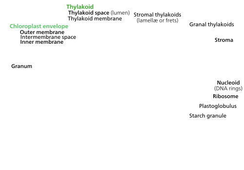
Chloroplast ultrastructure (interactive diagram) Chloroplasts have at least three distinct membrane systems, and a variety of things can be found in their stroma.
|


A mother and daughter centriole, attached orthogonally.
|
|


1.1 Porins
2.2 Peripheral space
3 Lamellæ
3.1 Inner membrane
3.12 Cristal membrane
3.2 Matrix
3.3 Cristæ
6 Ribosome
|
Mitochondrion ultrastructure (interactive diagram) A mitochondrion has a double membrane; the inner one contains its chemiosmotic apparatus and has deep grooves which increase its surface area. While commonly depicted as an "orange sausage with a blob inside of it" (like it is here), mitochondria can take many shapes and their intermembrane space is quite thin.
|
-en.svg)
Many of these are some of my largest illustrations, formatted as posters you might print out and put up on a classroom wall. These illustrations generally depict various cell processes to varying degrees of detail.


Some additional illustrations of cells doing various things.


A eukaryote with mitochondria engulfed a cyanobacterium in an event of serial primary endosymbiosis, creating a lineage of cells with both organelles. It is important to note that the cyanobacterial endosymbiont already had a double membrane—the phagosomal vacuole-derived membrane was lost.
 |
When chloroplasts are exposed to direct sunlight, they stack along the anticlinal cell walls to minimize exposure. In the dark they spread out in sheets along the periclinal walls to maximize light absorption.
|
Some poster-size illustrations depicting various biological processes. They are all purely vector, except for the plant cell types and the C4 poster which include embedded bitmaps in the form of the circular photographs presented alongside the illustration.


 |
Many C4 plants have their mesophyll cells and bundle sheath cells arranged radially around their leaf veins. The two types of cells contain different types of chloroplasts specialized for a particular part of photosynthesis.
|
Illustrations of whole organisms, ranging from a cyanobacterium to a general plant.

 |
Both chloroplasts and cyanobacteria have a double membrane, DNA, ribosomes, and thylakoids. Both the chloroplast and cyanobacterium depicted are idealized versions (the chloroplast is that of a higher plant)—a lot of diversity exists among chloroplasts and cyanobacteria.
|


These are a bunch of AP-level genetics posters illustrating some important genetics processes. Admittedly they contain quite too much text, and so they don’t really work that well. The DNA helices were rendered using a 3D app called Blender.


