Category:Anatomy of the human foot
Subcategories
This category has the following 15 subcategories, out of 15 total.
A
- Arches of the foot (3 F)
B
D
- Dissection of the human foot (27 F)
F
J
L
M
N
P
T
V
W
- Why the Shoe Pinches (15 F)
Media in category "Anatomy of the human foot"
The following 69 files are in this category, out of 69 total.
-
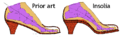 5,782,015 insolia.png 696 × 216; 7 KB
-

-
 Adult and foetal heel bone (calcaneus).jpg 1,942 × 3,281; 400 KB
-
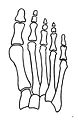
 Arch - Foot (PSF).png 2,003 × 1,256; 58 KB
-

-
 Blausen 0411 FootAnatomy eu.png 750 × 1,500; 636 KB
-
 Bones of the feet; two figures. Lithograph by Battistelli af Wellcome V0008194EL.jpg 1,149 × 1,548; 1,019 KB
-
 Bones of the feet; two figures. Lithograph by Battistelli af Wellcome V0008194ER.jpg 1,180 × 1,671; 1.07 MB
-
 Bones of the foot. Colour wood engraving with letterpress Wellcome V0008694.jpg 2,124 × 3,402; 3.5 MB
-
 Braus 1921 303.png 1,090 × 638; 1.99 MB
-
 Braus 1921 311.1.png 1,553 × 1,518; 6.76 MB
-
 Braus 1921 311.2.png 1,469 × 1,520; 6.4 MB
-
 Braus 1921 312.png 1,520 × 1,698; 7.4 MB
-
 Bänder am Fuss - 4686.jpg 2,081 × 4,720; 4.82 MB
-
 Bänder am Fuss - 4716.jpg 1,096 × 2,252; 1.07 MB
-
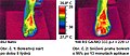
 Cévní obraz - srovnání před a po.jpg 763 × 726; 91 KB
-
-
 Dešinės pėdos jungtys.png 897 × 650; 475 KB
-
 Die Gartenlaube (1855) b 530.jpg 349 × 466; 33 KB
-
 Fasciaplantar-frontal.jpg 1,439 × 2,259; 1.3 MB
-
 First metatarsal bone has moved aside1.jpg 444 × 683; 50 KB
-
 Foot "Anatomia Humani Corporis", Bidloo, 1685 Wellcome L0014232.jpg 1,186 × 1,690; 759 KB
-
 Foot Arche (PSF).png 2,053 × 1,357; 348 KB
-

-

-
 Foot Chart1 small.png 249 × 314; 26 KB
-
 Foot chart1.png 554 × 697; 205 KB
-
 Foot of a 13 year old - 4688.jpg 747 × 1,818; 901 KB
-
 Foot retro.JPG 2,288 × 1,712; 1.35 MB
-
 Foot.png 214 × 356; 10 KB
-
 Gray1239 Arabic YM.jpg 3,496 × 3,912; 2.02 MB
-
 Hallux Romanux 2.jpg 1,920 × 2,560; 1.21 MB
-
 Hallux Romanux.jpg 1,920 × 2,560; 1.15 MB
-
 Homo footprints.jpg 1,552 × 928; 153 KB
-
 Huesos hallux.jpg 766 × 336; 39 KB
-
 Huesos hallux2.jpg 225 × 801; 55 KB
-
 MNH - Mumie Fuß.jpg 3,390 × 1,998; 1.41 MB
-
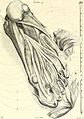
 Muscles- feet and hand Wellcome L0006624.jpg 1,136 × 1,751; 495 KB
-
 Nerfs de la face plantaire du pied droit.png 591 × 742; 166 KB
-
 Normal foot skeleton1.jpg 452 × 693; 52 KB
-
 NormaleFusssohle mit eingezeichbetemSkelett undBodenflache.gif 1,119 × 2,919; 127 KB
-
 Obr12.jpg 380 × 166; 117 KB
-
-
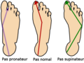 Pas pronateur-supinateur-normal.png 400 × 297; 40 KB
-
 PF-PainAreas-ar.jpg 742 × 875; 202 KB
-
 PF-PainAreas.jpg 742 × 875; 62 KB
-
 PF-PlantarMove.jpg 622 × 866; 55 KB
-
 Plantar Bewegung.gif 524 × 720; 54 KB
-
 Plantar Cutaneous Nerves.png 162 × 104; 36 KB
-
 Plantar fibromatosis.jpg 1,099 × 1,758; 274 KB
-
 Posicion referencia.JPG 768 × 576; 55 KB
-
 PSM V17 D758 Normal and boot deformed feet.jpg 3,806 × 2,869; 701 KB
-
 PSM V24 D664 Well formed healthy foot.jpg 1,008 × 1,585; 224 KB
-
 PSM V24 D666 Outlines of a normal foot and a cramping shoe.jpg 1,678 × 623; 105 KB
-
 Röntgen, Hammerzeh am mittleren Zeh.jpg 769 × 1,259; 203 KB
-
 Science ofDressTo face p239.png 1,493 × 3,072; 501 KB
-
 Science ofDressTo face p239cut.png 1,157 × 3,072; 360 KB
-
 Science ofDressTo face p240.png 2,000 × 3,072; 1.11 MB
-
 Science ofDressTo face p240cut.png 2,200 × 3,072; 1.02 MB
-
 Science ofDressTo face p240cutA.png 1,001 × 3,072; 769 KB
-
 Science ofDressTo face p240cutB.png 1,058 × 3,072; 738 KB
-
 Sehne Musculus extensor hallucis longus - Sono laengs zusammengesetzt - 001.png 1,155 × 291; 185 KB
-
 Slide2ALE.JPG 960 × 720; 80 KB
-
 Soldiersfootmili00munsrich Fig7 anteroposterior arch.png 1,012 × 572; 61 KB
-
 Text-book of operative surgery (1903) (14762066304).jpg 2,329 × 3,697; 1.4 MB
-
 The bones of the foot, from Camper, Dissertation ... Wellcome L0004215.jpg 1,280 × 1,452; 966 KB
-
 TPedBar6.jpg 200 × 396; 9 KB
-
 UBC-BME-KNT-20-008.png 756 × 588; 114 KB
-

_(14597413648).jpg)
.jpg)
.png)
_(14754215121).jpg)


_(14596371460).jpg)

_b_530.jpg)


.png)


_(14801787723).jpg)


_(14762066304).jpg)


_v_Praze_(ilustracni_fotografie)_(UK0242).jpg)