Category:Andy Lehrer
Romanian entomologist (1930-2014)  | |||||
| Upload media | |||||
| Date of birth | 16 May 1930 Iași | ||||
|---|---|---|---|---|---|
| Date of death | 6 February 2014 Israel | ||||
| Country of citizenship | |||||
| Occupation | |||||
| Employer |
| ||||
| |||||
Media in category "Andy Lehrer"
The following 58 files are in this category, out of 58 total.
-
 Abdomen de Sarcophaga carnaria.jpg 2,835 × 3,442; 2.64 MB
-
 Abdomenul la Calliphora.jpg 3,156 × 3,232; 1.37 MB
-
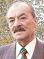 Andy Lehrer.jpg 158 × 211; 11 KB
-
 Bengalia.jpg 686 × 463; 84 KB
-
 Bengaliidae Lehrer n. fam..pdf 1,020 × 1,387, 10 pages; 589 KB
-
 Chetotaxia capului la Calliphora.jpg 3,706 × 4,156; 2.81 MB
-
 Chetotaxie de la tête du mâle.gif 2,835 × 2,777; 159 KB
-
 Chétotaxie de la tete de la femelle.jpg 2,835 × 2,740; 1.04 MB
-
 Chétotaxie de la tete du male.jpg 2,835 × 2,778; 1.33 MB
-
 Chétotaxie de la tête de la femelle.gif 2,835 × 2,740; 131 KB
-
 Chétotaxie du thorax dorsal.gif 2,835 × 3,276; 169 KB
-
 Chétotaxie du thorax latéral.gif 3,072 × 2,217; 116 KB
-
 Chétotaxie du thorax, vu dorsal.jpg 2,835 × 3,277; 1.43 MB
-
 Chétotaxie du thorax, vu lateral.jpg 2,835 × 2,047; 879 KB
-
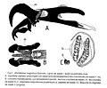
 Falozomul speciei Afridigalia adrianponti.jpg 2,494 × 2,818; 1.48 MB
-
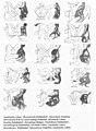
 Fig. 1. Bengalia labiata Robineau-Desvoidy.jpg 3,320 × 2,762; 1.46 MB
-
 Fig. 10. Abdomen de Sarcophaga carnaria.jpg 2,363 × 3,171; 1.02 MB
-
 Fig. 2. Larve de stade III de Wohlfahrtia sp..jpg 1,418 × 847; 251 KB
-
 Fig. 3. Gangelomyia xanthopyga SW.jpg 3,582 × 2,544; 1.59 MB
-
 Fig. 32. Afridigalia tibiaria Vill.jpg 3,460 × 3,118; 1.84 MB
-
 Fig. 35.Kenypyga bantuphalla Lehrer n.jpg 3,970 × 3,148; 1.91 MB
-
 Fig. 4. Maraviola spinifemorata Vill.jpg 3,574 × 3,410; 1.52 MB
-
 Fig. 6. Podomyiase et ophthalmomyiase.jpg 2,835 × 1,496; 1.7 MB
-
 Fig. 7. Types de structure sfam copy.gif 2,425 × 3,929; 278 KB
-
 Genitalia de l'Afridigalia gaillardi.jpg 3,802 × 2,776; 1.21 MB
-
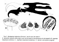
 Genitalia speciei Afridigalia adrianponti Lhrr.jpg 3,214 × 3,126; 2 MB
-
 Genitalia speciei Ashokiana ramsdalei Lhrr.jpg 3,610 × 2,858; 1.93 MB
-
 Genitalia speciei Bengalia fernandiella Lhrr. 2005.jpg 3,078 × 2,676; 1.52 MB
-
 Genitalia speciei Bengalia lampunia Lhrr. 2005 .jpg 2,756 × 3,058; 1.51 MB
-
 Genitalia speciei Bengalia nirvanella Lhrr 2005.jpg 3,780 × 2,892; 1.73 MB
-
 Genitalia speciei Gangelomyia krishna Lhrr. 2005.jpg 3,730 × 2,802; 1.9 MB
-
 Genitalia speciei Hyperacanthisca susteriana Lehrer, 2003.jpg 1,418 × 946; 293 KB
-
 Genitalia speciei Sarcophaga susteri Lehrer.jpg 2,702 × 1,962; 1.23 MB
-
 Genres des Sarcophagidae.jpg 3,709 × 5,054; 4.67 MB
-
 Larve de stade II-B.jpg 2,985 × 2,581; 644 KB
-
 Larve de stade II.jpg 2,952 × 2,137; 516 KB
-
 Larve de stade III de Sarcophaginae.jpg 1,418 × 652; 120 KB
-
 Larve de stade III.jpg 2,965 × 2,281; 901 KB
-
 Larve I Wohlfahrtia.jpg 1,418 × 1,161; 270 KB
-
 Lave de stade I-B.jpg 3,079 × 2,534; 907 KB
-
 Lave de stade I.jpg 2,858 × 3,108; 1.04 MB
-
 Myiases génitales graves.jpg 2,835 × 2,101; 2.59 MB
-
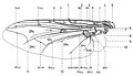 Nervation des Sarcophagidae.jpg 2,835 × 1,620; 547 KB
-
 Nervaţiunea aripii la Calliphora.jpg 3,138 × 2,630; 1.56 MB
-
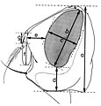 Parametres dimensionnels de la tete, vu de profil..jpg 2,835 × 2,966; 1.33 MB
-
 Parametres dimensionnels de la tete, vu du dessus.jpg 2,835 × 2,823; 1.51 MB
-
 Phallosome de Sarcophaga carnaria.jpg 2,835 × 3,771; 1.67 MB
-
 Postabdomen de Sarcophaga carnaria.jpg 2,835 × 1,147; 423 KB
-
 Postabdomen de Sarcophaga.jpg 2,835 × 1,506; 630 KB
-
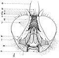
 Postabdomenul la Calliphora.jpg 3,240 × 3,992; 1.39 MB
-
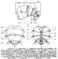
 Regiunile si zonele capului la mascul.jpg 3,106 × 1,942; 780 KB
-
 Sarcophaga carnaria Böttcher.jpg 219 × 326; 16 KB
-
 Shakaniella wyatti Lehrer. 2005.jpg 3,406 × 3,214; 1.86 MB
-
 Squelette cephalo-pharyngeal III.jpg 1,418 × 701; 107 KB
-
 Sternites postabdominaux VI et VII des Bengaliidae.jpg 3,476 × 2,382; 1.68 MB
-
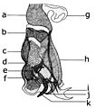
 Structura falozomului la Calliphora.jpg 3,096 × 2,240; 1.07 MB
-
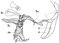
 Toracele la Calliphora.jpg 3,076 × 3,134; 1.4 MB
-
 Wohlfahtiose vulvaire.jpg 1,604 × 1,217; 1.6 MB


