Category:Bones of the human skull
 Human anatomy | ||
|---|---|---|
Subcategories
This category has the following 23 subcategories, out of 23 total.
Pages in category "Bones of the human skull"
This category contains only the following page.
Media in category "Bones of the human skull"
The following 101 files are in this category, out of 101 total.
-
 0.4.MHNT.ANT.1996.04 Crane Humaine éclaté à la Beauchêne.jpg 4,268 × 4,489; 5.35 MB
-
 3381250812 75bb40a1d3 oCraneEclaté.jpg 533 × 789; 263 KB
-
 730 Posterior View Skull.jpg 810 × 631; 201 KB
-
 Apatine nosies kriaukle.JPG 1,398 × 822; 181 KB
-
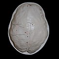
 Base of cranium (11291052176).jpg 1,475 × 2,076; 831 KB
-
 Base of cranium (11291131743).jpg 1,632 × 2,464; 1.29 MB
-
 Base of cranium and nose (11291003395).jpg 2,210 × 1,480; 682 KB
-
 Base of skull 1.jpg 960 × 720; 116 KB
-
 Base of skull 10.jpg 960 × 720; 115 KB
-
 Base of skull 13.jpg 960 × 720; 105 KB
-
 Base of skull 15.jpg 800 × 600; 207 KB
-
 Base of skull 16.jpg 960 × 720; 107 KB
-
 Base of skull 19-ar.jpg 960 × 720; 350 KB
-
 Base of skull 19.jpg 960 × 720; 107 KB
-
 Base of skull 24.jpg 960 × 720; 71 KB
-
 Base of skull 4.jpg 960 × 720; 105 KB
-
 Base of skull 5.jpg 960 × 720; 103 KB
-
 Base of skull 6.jpg 960 × 720; 112 KB
-
 Base of skull 7.jpg 960 × 720; 112 KB
-
 Base of skull 8.jpg 960 × 720; 113 KB
-
 Base of skull 9.jpg 960 × 720; 113 KB
-
 Braus 1921 334.png 1,476 × 944; 3.99 MB
-
 Cavitatea bucală.jpg 500 × 507; 72 KB
-
 Child skull.jpg 960 × 720; 74 KB
-
 Child viscerocranium 1.jpg 960 × 720; 65 KB
-
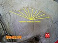
 Coronal suture 2.jpg 960 × 720; 59 KB
-
 Coronal suture.jpg 960 × 720; 65 KB
-
 Crane-img 0513.jpg 2,048 × 3,072; 418 KB
-
 Crane-img 0514.jpg 2,048 × 3,072; 547 KB
-
 Crane-img 0515.jpg 2,048 × 3,072; 569 KB
-
 Crane4 Foramen magnum.png 2,431 × 2,761; 6.83 MB
-
 Crane4.png 2,431 × 2,761; 6.96 MB
-
 Cranio. Fossa pterigo-palatina.jpg 960 × 720; 96 KB
-
 Cranio. Norma laterale. Fossa Temporale.jpg 960 × 720; 103 KB
-
 Cranio. Palato osseo.jpg 960 × 720; 69 KB
-
 Cranio. Punti craniometrici.jpg 960 × 720; 50 KB
-
 Cranio. Suture dello splancnocranio.jpg 960 × 720; 90 KB
-
 Cranium and nose (11291075594).jpg 1,571 × 1,389; 464 KB
-
 Cranium, nose and eye (11291125153).jpg 2,464 × 1,632; 782 KB
-
 Ejemplo frecuente de CAT (1).jpg 172 × 151; 4 KB
-
 Ejemplo frecuente de CAT (2).jpg 170 × 191; 6 KB
-
 Ejemplo frecuente de CAT (3).jpg 168 × 203; 5 KB
-
 Ejemplo frecuente de CAT (4).jpg 227 × 170; 5 KB
-
 Endobasis - resistances beams.jpg 960 × 720; 97 KB
-
 Endobasis - resistances nodes.jpg 960 × 720; 111 KB
-
 Endocranium.jpg 1,517 × 1,801; 1.24 MB
-
 EsztregomFotoThaler4.jpg 1,024 × 683; 187 KB
-
 Exobasis.jpg 960 × 720; 62 KB
-
 Exploded Human Skull.jpg 1,349 × 1,457; 265 KB
-
 Fetal skull11w.jpg 584 × 531; 39 KB
-
 Foramen ethmoidale anterius.PNG 719 × 1,057; 149 KB
-
 Foramen ethmoidale posterius.PNG 719 × 1,057; 149 KB
-
 G.F. Ingrassia, "In Galeni librum de ossibus..." Wellcome L0028114.jpg 1,154 × 1,786; 1.11 MB
-
 Gray189 zh.png 500 × 503; 420 KB
-
 Gray189.png 500 × 503; 387 KB
-
 Gray194 zh.png 650 × 420; 245 KB
-
 Gray194.png 650 × 420; 84 KB
-
 Human palatine bone (left).png 747 × 859; 312 KB
-
 Human skull 01.jpg 3,872 × 2,592; 4.13 MB
-
 Human skull side suturas right-IT.png 612 × 396; 119 KB
-
 Internal bones of nose, in Iconum anatomicarum. Wellcome M0017123.jpg 4,134 × 2,725; 2.41 MB
-
 Lambdoid suture.jpg 960 × 720; 62 KB
-
 Lectures on the elements of comparative anatomy (Page 119) BHL3318924.jpg 2,326 × 3,749; 728 KB
-
 Lectures on the elements of comparative anatomy (Page 120) BHL3318839.jpg 2,316 × 3,759; 659 KB
-
 Lectures on the elements of comparative anatomy (Page 158) BHL3318834.jpg 2,316 × 3,759; 629 KB
-
 Merkel's Human Anatomy (1913) - Vol 3 - Fig 045.png 1,344 × 1,204; 660 KB
-
 Merkel's Human Anatomy (1913) - Vol 3 - Fig 046.png 1,128 × 1,227; 570 KB
-
 Neumarkt-an-der-Ybbs-Grab-74-Dünnschliff-des-Schädelknochens.jpg 1,833 × 816; 1.08 MB
-
 NNH im Roentgen frontal.png 678 × 962; 393 KB
-
 Nosikaulis.jpg 200 × 230; 21 KB
-
 Oberlin-Oreille.jpg 2,328 × 1,818; 569 KB
-
 Occipital bone.jpg 960 × 720; 76 KB
-
 Orbita mensch.jpg 500 × 374; 64 KB
-
 Orbital bones.png 350 × 350; 104 KB
-
 Os frontal1.png 692 × 745; 255 KB
-
 Palate (PSF).png 3,240 × 1,384; 288 KB
-
 Palate 2 (PSF).png 1,341 × 1,238; 178 KB
-
 Sagittal suture 2.jpg 960 × 720; 71 KB
-
 Sagittal suture 3.jpg 960 × 720; 60 KB
-
 Sagittal suture.jpg 960 × 720; 71 KB
-
 Schaedel im Röntgen seitlich.png 1,092 × 1,078; 640 KB
-
 Schädelbasis1.jpg 1,517 × 1,801; 1.17 MB
-
 Skull - midsaggital section P.2005.jpg 1,024 × 937; 62 KB
-
 Skullfront.png 1,220 × 1,872; 1.81 MB
-
 SkullSchaedel1.png 900 × 1,200; 36 KB
-
 SkullSchaedel2.png 900 × 1,200; 40 KB
-
 SkullSchaedel3.png 900 × 1,200; 36 KB
-
 Slide3ROM-ar.JPG 824 × 600; 258 KB
-
 Slide3ROM.JPG 960 × 720; 84 KB
-
 Slide4gre.JPG 960 × 720; 74 KB
-
 Slide6gre.JPG 960 × 720; 88 KB
-
 Sphenoid bone with anterior clinoid processes highlighted.png 1,183 × 883; 653 KB
-
 Temporo-parietal suture 3.jpg 960 × 720; 93 KB
-
 Temporo-parietal suture 4.jpg 960 × 720; 73 KB
-
 Veido kaulai.JPG 663 × 850; 132 KB
-
 Vomer bone.png 800 × 455; 314 KB
-
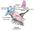 Vomer.png 635 × 572; 143 KB
-
 Vomer1.png 564 × 427; 34 KB
-
 Θόλος του κρανίου ..jpg 4,858 × 4,858; 2.81 MB
-
 Θόλος του κρανίου.jpg 4,858 × 4,858; 2.78 MB
-
 Κρανίο.jpg 4,000 × 4,000; 2.61 MB


.jpg)
.jpg)
.jpg)


.jpg)
.jpg)
.jpg)
.jpg)
.jpg)
.jpg)


.png)


_BHL3318924.jpg)
_BHL3318839.jpg)
_BHL3318834.jpg)
_-_Vol_3_-_Fig_045.png)
_-_Vol_3_-_Fig_046.png)


.png)
.png)


