Category:Branchiostoma anatomy
Subcategories
This category has only the following subcategory.
Media in category "Branchiostoma anatomy"
The following 55 files are in this category, out of 55 total.
-
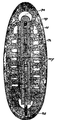
 1911 Britannica - Amphioxus lauceolatus.png 1,067 × 1,991; 285 KB
-
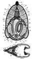
 1911 Britannica - Diagram of embryo of Amphioxus.png 516 × 1,089; 82 KB
-
 1911 Britannica - Pelagic larvae of A. lanceolatus.png 1,049 × 667; 63 KB
-
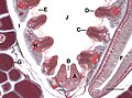
 1911 Britannica - Transverse sections of Amphioxus.png 1,086 × 1,891; 217 KB
-
 Amphioxus - Pharyngeal Region c.s..jpg 720 × 960; 86 KB
-
 Amphioxus bottom pharyngeal CS L.jpg 1,200 × 886; 143 KB
-
 Amphioxus bottom pharyngeal CS.jpg 1,200 × 886; 139 KB
-
 Amphioxus bottom pharyngeal zoom CS L.jpg 1,200 × 886; 194 KB
-
 Amphioxus bottom pharyngeal zoom CS.jpg 1,200 × 886; 193 KB
-
 Amphioxus cs Pharyngeal.jpg 1,179 × 1,528; 239 KB
-
 Amphioxus male CS L.jpg 787 × 1,494; 164 KB
-
 Amphioxus male CS.jpg 787 × 1,494; 164 KB
-
 Amphioxus pharyngeal CS L.jpg 1,027 × 1,200; 115 KB
-
 Amphioxus pharyngeal CS.jpg 1,027 × 1,200; 113 KB
-
 Amphioxus pharyngeal tail CS.jpg 1,026 × 1,200; 98 KB
-
 Amphioxus pharyngeal top CS L.jpg 1,200 × 886; 137 KB
-
 Amphioxus pharyngeal top CS.jpg 1,200 × 886; 133 KB
-
 Amphioxus pharyngeal top zoom CS L.jpg 1,200 × 886; 171 KB
-
 Amphioxus pharyngeal top zoom CS.jpg 1,200 × 886; 169 KB
-
 Amphioxus pharyngeal.jpg 1,200 × 886; 112 KB
-
 Amphioxus tail L.jpg 1,200 × 886; 70 KB
-
 Amphioxus tail.jpg 1,200 × 886; 67 KB
-
 Amphioxus whole L.jpg 1,200 × 628; 43 KB
-
 Amphioxus whole.jpg 1,200 × 628; 33 KB
-
 Animal biology (1938) (17574048064).jpg 3,200 × 1,278; 668 KB
-
 Anterior External L.jpg 1,075 × 1,445; 326 KB
-
 Branchiostoma cultellus1.jpg 1,000 × 768; 252 KB
-
 Branchiostoma lanceolatum (Pallas, 1774) - coupes transversales légendées.jpg 2,520 × 1,876; 3.86 MB
-
 Branchiostoma lanceolatum (Pallas, 1774) - coupes transversales.jpg 2,520 × 1,876; 3.73 MB
-
 Branchiostoma lanceolatum (Pallas, 1774) 1 - légendée.jpg 4,288 × 2,247; 3.17 MB
-
 Branchiostoma lanceolatum (Pallas, 1774) 3.jpg 3,620 × 1,344; 2.54 MB
-
 BranchiostomaLanceolatum PioM (hy).png 2,165 × 704; 286 KB
-
 Cepl003b.png 1,411 × 1,662; 182 KB
-
 Cepl013c.png 2,330 × 937; 484 KB
-
 Chor Cepl amphioxus head.jpg 2,048 × 1,536; 397 KB
-
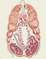
 Cross section of Amphioxus.jpg 800 × 598; 74 KB
-
 Gonadal Section L.jpg 1,666 × 1,220; 379 KB
-
 Lancelet GFP GIF.gif 600 × 600; 4.07 MB
-
 Lancelet's endostyle1, numbered.jpg 950 × 1,200; 402 KB
-
 Lancelet's endostyle1.jpg 950 × 1,200; 444 KB
-
 Lancelet's endostyle2, numbered.jpg 950 × 1,200; 512 KB
-
 Lancelet's endostyle2.jpg 950 × 1,200; 583 KB
-
 Lancelet's gill slits, numbered.jpg 1,200 × 950; 455 KB
-
 Lancelet's gill slits.jpg 1,200 × 950; 508 KB
-
 Lancelet's metapleural fold.jpg 1,200 × 950; 356 KB
-
 Lancelet's mid-gut diverticulum.jpg 950 × 1,600; 801 KB
-
 Lancelet's nerve cord.jpg 1,200 × 950; 620 KB
-
 Lancelet's ovary.jpg 950 × 1,200; 383 KB
-
 Lectures on the elements of comparative anatomy (Page 190) BHL3318940.jpg 2,316 × 3,759; 738 KB
-
 Meyers b5 s0350 b1.png 570 × 218; 53 KB
-
 Tail Section CS L.jpg 1,491 × 1,198; 189 KB
-
 The Amphioxus and its development (1893) (17542538974).jpg 1,846 × 2,740; 1.2 MB
-
 The Amphioxus and its development (1893) (17544525153).jpg 1,776 × 2,738; 1.39 MB
-
 The Amphioxus and its development (1893) (17978889339).jpg 2,624 × 1,710; 1.2 MB
-
 Transverse section of lancelet.jpg 950 × 1,200; 287 KB


_1_-_l%C3%A9gend%C3%A9e.jpg)


_BHL3318940.jpg)

_(17542538974).jpg)
_(17544525153).jpg)
_(17978889339).jpg)
_(17574048064).jpg)
_-_coupes_transversales_l%C3%A9gend%C3%A9es.jpg)
_-_coupes_transversales.jpg)
_3.jpg)
.png)