Category:Epiblast
Media in category "Epiblast"
The following 67 files are in this category, out of 67 total.
-
 2908 Germ Layers-02-nltxt.jpg 1,854 × 1,658; 1.45 MB
-
 2908 Germ Layers-02.jpg 1,781 × 1,590; 1.06 MB
-

-
 Agelena labyrinthica ventral plate at three stages.jpg 1,120 × 719; 454 KB
-

-
 Amphibia presumptive organ-forming areas in the late blastula and beginning gastrula.jpg 1,164 × 1,581; 1.61 MB
-
-

-
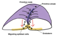
 Aves Clustering of seemingly stochastic EMT underpins the formation of PS.jpg 780 × 1,500; 174 KB
-
 Aves delamination of hypoblast (entoderm) cells from upper or epiblast layer.jpg 1,093 × 429; 575 KB
-
 Aves Diagrams depicting the early stages of chick development.jpg 1,500 × 922; 152 KB
-
-
 Aves gastrulation Tissue flows driving primitive streak formation.jpg 1,663 × 1,621; 710 KB
-
 Aves origin of the hypoblast (entoderm) in the avian blastoder.jpg 995 × 507; 484 KB
-
 Avian development before gastrulation.jpg 2,767 × 3,354; 1.41 MB
-
-
 Cat longitudinal section through the axis of the ovum.jpg 878 × 706; 839 KB
-
 Cat Yolk segmentation.jpg 1,202 × 470; 563 KB
-

-
 Characterization of the SOX2T-positive territory of the epiblast in chicken embryo.jpg 2,113 × 1,581; 1.21 MB
-
 Chick embryo. longitudinal section of a chick of the fourth day.jpg 1,344 × 534; 307 KB
-
 Cilia are present and functional in the node of 4HH-8HH talpid3 chickens.jpg 1,063 × 1,402; 582 KB
-
-
 Didelphidae early development of blastoderm.jpg 1,045 × 788; 621 KB
-
 Different types of EMT.jpg 1,233 × 935; 179 KB
-
 Dose-dependent reshaping of primitive streak.jpg 968 × 1,236; 743 KB
-
Dynamics of mesodermal cell ingression chicken embryo.ogv 12 s, 226 × 720; 4.2 MB
-

-
 EB1911 Tunicata - Stages in the Embryology of a Simple Ascidian.jpg 805 × 913; 216 KB
-

-
 EmbryonVitellinPrimaire.jpg 1,676 × 2,025; 735 KB
-
 Euaxes ovum during early stage of development.jpg 876 × 631; 547 KB
-
-
-
 Fundulus heteroclitus presumptive organ-forming areas of the blastoderm.jpg 1,020 × 796; 779 KB
-
 Hand-book of physiology (1892) (14742413116).jpg 1,888 × 680; 245 KB
-
 Hand-book of physiology (1892) (14765102622).jpg 1,188 × 308; 375 KB
-
 Holothuria tubulosa development embryo.jpg 1,224 × 675; 776 KB
-
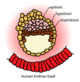 Human Embryo Day9.png 323 × 330; 54 KB
-

-
 Hydrophilus piceus embryos (01).jpg 754 × 750; 520 KB
-

-
Live imaging of Histone H2B–GFP mouse embryo at E6.5.ogv 6.4 s, 804 × 631; 674 KB
-
Live imaging of mouse embryo at E5.5.ogv 4.3 s, 366 × 375; 145 KB
-
Live imaging of mouse embryo at E6.ogv 8.6 s, 520 × 491; 497 KB
-
 Live imaging of the whole mouse embryo at E6 (A to D) and E5.5. (E to H).png 1,910 × 2,240; 3.3 MB
-
 Live imaging of the whole mouse embryo at E6.5 using DSLM.png 2,059 × 1,366; 2.24 MB
-
Longevity of tracks along the primitive streak (PS) chicken embryo.ogv 5.9 s, 286 × 720; 1.48 MB
-
 Migration of epiblast cells in the mammalian embryo.png 866 × 530; 281 KB
-
 Quantification of the number of epiblast cells electroporated chicken embryo.jpg 730 × 1,687; 273 KB
-
 Rana temporaria cleavage at the close of segmentation embryo.jpg 768 × 727; 627 KB
-
Reconstructed epiblast nuclei from a lateral view.ogv 6.4 s, 1,031 × 728; 518 KB
-
Reconstructed epiblast nuclei from distal view.ogv 6.4 s, 924 × 636; 571 KB
-
Reconstructed trajectories of mesodermal cells.ogv 4.3 s, 637 × 567; 807 KB
-
 Reptilia formation of hypoblast (entoderm) layer.jpg 961 × 1,101; 1.03 MB
-
 Salmo irideus presumptive organ-forming areas in the blastoderm.jpg 1,163 × 884; 685 KB
-

-
 Scorpion embryo mesoblastic somites.jpg 527 × 736; 354 KB
-
 Selachimorpha presumptive organ-forming areas in the blastoderm.jpg 981 × 801; 617 KB
-
 Simiiformes developing blastocyst.jpg 1,105 × 1,369; 668 KB
-
 Sus domesticus blastocyst.jpg 1,016 × 759; 647 KB
-

-
Time-lapse changes of triangles connected neighboring nuclei at the first time point (2).ogv 5.5 s, 638 × 541; 1.77 MB
-
Time-lapse migrations of mesodermal cells.ogv 9.1 s, 635 × 569; 2.26 MB
-
Tracking descendants of the anterior primitive streak (PS) epiblast at 25 somites chicken embryo.ogv 30 s, 444 × 720; 10.99 MB
-
Tracking the fate of descendants of the anterior epiblast at 25 somites chicken embryo.ogv 12 s, 1,280 × 578; 6.39 MB
-


_(14596981329).jpg)


_in_the_avian_blastoder.jpg)


_(20670956325).jpg)


.jpg)

_and_E5.5._(E_to_H).png)


_layer.jpg)

_and_median_sagittal_sections_(g_and_h)_using_the_rabbit_as_a_model.jpg)


_(20398797108).jpg)
_cells_from_upper_or_epiblast_layer.jpg)
_(14742413116).jpg)
_(14765102622).jpg)