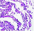
Category:Histopathology of adenocarcinomas of the human lung
Media in category "Histopathology of adenocarcinomas of the human lung"
The following 113 files are in this category, out of 113 total.
-
Acinar pattern adenocarcinoma of lung - alt -- very high mag.jpg 4,272 × 2,848; 4.2 MB
-
Acinar pattern adenocarcinoma of lung -- high mag.jpg 4,272 × 2,848; 4.66 MB
-
Acinar pattern adenocarcinoma of lung -- intermed mag.jpg 4,272 × 2,848; 5.06 MB
-
Acinar pattern adenocarcinoma of lung -- low mag.jpg 4,272 × 2,848; 3.65 MB
-
Acinar pattern adenocarcinoma of lung -- very high mag.jpg 4,272 × 2,848; 4.19 MB
-
Adenocarcinoma - "Mucinous pneumonia" (5496448423).jpg 2,272 × 1,704; 1.59 MB
-
Adenocarcinoma - "Mucinous pneumonia" (5497041416).jpg 2,272 × 1,704; 1.58 MB
-
Adenocarcinoma - Napsin A immunostain (5762462036).jpg 2,048 × 1,536; 1.18 MB
-
Adenocarcinoma - Napsin A immunostain (6838767882).jpg 2,048 × 1,536; 1.05 MB
-
Adenocarcinoma - ROS1 positive - napsin A -- high mag.jpg 6,000 × 4,000; 9.21 MB
-
Adenocarcinoma - ROS1 positive - napsin A -- intermed mag.jpg 6,000 × 4,000; 8.94 MB
-
Adenocarcinoma - ROS1 positive - napsin A -- very high mag.jpg 6,000 × 4,000; 7.59 MB
-
Adenocarcinoma - ROS1 positive - ROS1 -- high mag.jpg 6,000 × 4,000; 7.6 MB
-
Adenocarcinoma - ROS1 positive - ROS1 -- intermed mag.jpg 6,000 × 4,000; 8.12 MB
-
Adenocarcinoma - ROS1 positive - ROS1 -- very high mag.jpg 6,000 × 4,000; 6.96 MB
-
Adenocarcinoma - ROS1 positive - TTF1 -- high mag.jpg 6,000 × 4,000; 7.83 MB
-
Adenocarcinoma - ROS1 positive - TTF1 -- intermed mag.jpg 6,000 × 4,000; 6.66 MB
-
Adenocarcinoma - ROS1 positive - TTF1 -- very high mag.jpg 6,000 × 4,000; 5.71 MB
-
Adenocarcinoma - ROS1 positive -- high mag.jpg 6,000 × 4,000; 7.84 MB
-
Adenocarcinoma - ROS1 positive -- intermed mag.jpg 6,000 × 4,000; 8.99 MB
-
Adenocarcinoma - ROS1 positive -- low mag.jpg 6,000 × 4,000; 10.43 MB
-
Adenocarcinoma - ROS1 positive -- very high mag.jpg 6,000 × 4,000; 7.3 MB
-
Adenocarcinoma in situ, non-mucinous (9280005090).jpg 2,272 × 1,704; 2.65 MB
-
Adenocarcinoma of lung, FNA (5435902375).jpg 1,634 × 1,090; 825 KB
-
Adenocarcinoma with bronchiolar involvement (5762349504).jpg 2,048 × 1,536; 1.25 MB
-
Adenocarcinoma with clear cells (4420421470).jpg 1,600 × 1,200; 702 KB
-
Adenocarcinoma with clear cells (5496433011).jpg 1,479 × 1,199; 1.13 MB
-
Adenocarcinoma with clear cells (5497026036).jpg 1,482 × 1,199; 1.23 MB
-
Adenocarcinoma with sarcomatoid change - lung -- high mag.jpg 4,272 × 2,848; 5.64 MB
-
Adenocarcinoma with sarcomatoid change - lung -- intermed mag.jpg 4,272 × 2,848; 5.86 MB
-
Adenocarcinoma with sarcomatoid change - lung -- very high mag.jpg 4,272 × 2,848; 4.71 MB
-
Adenocarcinoma, acinar subtype, with lepidic growth at the periphery (7145417241).jpg 2,048 × 1,536; 3.25 MB
-
ALK positive lung adenocarcinoma - ALK IHC - alt -- high mag.jpg 4,000 × 6,000; 9.01 MB
-
ALK positive lung adenocarcinoma - ALK IHC - alt -- very high mag.jpg 4,000 × 6,000; 9.88 MB
-
ALK positive lung adenocarcinoma - ALK IHC -- high mag.jpg 4,000 × 6,000; 14.94 MB
-
ALK positive lung adenocarcinoma - ALK IHC -- intermed mag.jpg 4,000 × 6,000; 12.13 MB
-
ALK positive lung adenocarcinoma - ALK IHC -- low mag.jpg 4,000 × 6,000; 6.38 MB
-
ALK positive lung adenocarcinoma - ALK IHC -- very high mag.jpg 4,000 × 6,000; 10.19 MB
-
ALK positive lung adenocarcinoma - ALK IHC -- very low mag.jpg 4,000 × 6,000; 4.02 MB
-
ALK positive lung adenocarcinoma - alt -- high mag.jpg 4,000 × 6,000; 9.7 MB
-
ALK positive lung adenocarcinoma -- high mag.jpg 4,000 × 6,000; 15.01 MB
-
ALK positive lung adenocarcinoma -- intermed mag.jpg 4,000 × 6,000; 13.55 MB
-
ALK positive lung adenocarcinoma -- low mag.jpg 4,000 × 6,000; 6.53 MB
-
ALK positive lung adenocarcinoma -- very high mag.jpg 4,000 × 6,000; 10.67 MB
-
ALK positive lung adenocarcinoma -- very low mag.jpg 4,000 × 6,000; 4.67 MB
-
Clear cell adenocarcinoma of the lung.jpg 797 × 512; 382 KB
-
Fetal adenocarcinoma of the lung - alt1 -- very low mag.jpg 6,000 × 4,000; 16.95 MB
-
Fetal adenocarcinoma of the lung - alt2 -- very low mag.jpg 6,000 × 4,000; 16.58 MB
-
Fetal adenocarcinoma of the lung -- high mag.jpg 6,000 × 4,000; 15.49 MB
-
Fetal adenocarcinoma of the lung -- intermed mag.jpg 6,000 × 4,000; 17.79 MB
-
Fetal adenocarcinoma of the lung -- low mag.jpg 6,000 × 4,000; 16.07 MB
-
Fetal adenocarcinoma of the lung -- very low mag.jpg 6,000 × 4,000; 16.34 MB
-
Histopathology of lepidic predominant adenocarcinoma.jpg 531 × 488; 119 KB
-
Histopathology of lung adenocarcinoma with acinar pattern.png 702 × 705; 1.06 MB
-
Histopathology of lung adenocarcinoma with solid pattern.jpg 468 × 413; 128 KB
-
Intra-alveolar growth of poorly differentiated adenocarcinoma (5600778203).jpg 2,560 × 1,920; 2.05 MB
-
Intra-alveolar growth of poorly differentiated adenocarcinoma (5601362364).jpg 2,560 × 1,920; 2.52 MB
-
Invasive mucinous adenocarcinoma (8207250938).jpg 3,264 × 2,448; 2.9 MB
-
Lung adenocarcinoma - TTF-1 - high mag.jpg 4,272 × 2,848; 6.04 MB
-
Lung adenocarcinoma visceral pleural invasion - alt -- intermed mag.jpg 2,848 × 4,272; 5.65 MB
-
Lung adenocarcinoma visceral pleural invasion - Miller - alt -- high mag.jpg 2,848 × 4,272; 6.61 MB
-
Lung adenocarcinoma visceral pleural invasion - Miller - alt -- intermed mag.jpg 2,848 × 4,272; 6.35 MB
-
Lung adenocarcinoma visceral pleural invasion - Miller -- high mag.jpg 4,272 × 2,848; 5.39 MB
-
Lung adenocarcinoma visceral pleural invasion - Miller -- intermed mag.jpg 4,272 × 2,848; 6.11 MB
-
Lung adenocarcinoma visceral pleural invasion - Miller -- low mag.jpg 4,272 × 2,848; 5.64 MB
-
Lung adenocarcinoma visceral pleural invasion - Miller -- very high mag.jpg 2,848 × 4,272; 5.35 MB
-
Lung adenocarcinoma visceral pleural invasion - Miller -- very low mag.jpg 4,272 × 2,848; 5.75 MB
-
Lung adenocarcinoma visceral pleural invasion -- high mag.jpg 2,848 × 4,272; 4.72 MB
-
Lung adenocarcinoma visceral pleural invasion -- intermed mag.jpg 4,272 × 2,848; 5.44 MB
-
Lung adenocarcinoma visceral pleural invasion -- low mag.jpg 4,272 × 2,848; 5.05 MB
-
Lung adenocarcinoma visceral pleural invasion -- very high mag.jpg 2,848 × 4,272; 4.29 MB
-
Lung adenocarcinoma visceral pleural invasion -- very low mag.jpg 4,272 × 2,848; 5.28 MB
-
Lung adenocarcinoma with lepidic growth - low magnification 2.jpg 1,734 × 1,156; 878 KB
-
Lung adenocarcinoma with lepidic growth - low magnification.jpg 1,738 × 1,158; 1,015 KB
-
Metastatic endometrial adenocarcinoma, intrabronchial (8533050401).jpg 2,560 × 1,920; 2.85 MB
-
Mucinous adenocarcinoma (7145439953).jpg 1,600 × 1,200; 793 KB
-
Mucinous adenocarcinoma of the lung -- high mag.jpg 4,272 × 2,848; 4.83 MB
-
Mucinous adenocarcinoma of the lung -- intermed mag.jpg 4,272 × 2,848; 3.7 MB
-
Mucinous adenocarcinoma of the lung -- low mag.jpg 4,272 × 2,848; 3.07 MB
-
Mucinous adenocarcinoma of the lung -- very high mag.jpg 4,272 × 2,848; 4.38 MB
-
Mucinous lung adenocarcinoma - alt -- intermed mag.jpg 4,272 × 2,848; 4.58 MB
-
Mucinous lung adenocarcinoma -- high mag.jpg 4,272 × 2,848; 4.38 MB
-
Mucinous lung adenocarcinoma -- intermed mag.jpg 4,272 × 2,848; 4.78 MB
-
Mucinous lung adenocarcinoma -- low mag.jpg 4,272 × 2,848; 5.46 MB
-
Mucinous lung adenocarcinoma -- very high mag.jpg 4,272 × 2,848; 4.09 MB
-
Mucinous lung adenocarcinoma -- very low mag.jpg 4,272 × 2,848; 5.87 MB
-
Mucinous lung adenocarcinoma and airway - alt -- high mag.jpg 4,272 × 2,848; 4.95 MB
-
Mucinous lung adenocarcinoma and airway -- high mag.jpg 4,272 × 2,848; 4.97 MB
-
Mucinous lung adenocarcinoma and airway -- intermed mag.jpg 4,272 × 2,848; 4.92 MB
-
Neural invasion by pulmonary adenocarcinoma in mediastinum (7307509568).jpg 1,600 × 1,200; 744 KB
-
Papillary adenocarcinoma of the lung -- high mag.jpg 4,272 × 2,848; 4.86 MB
-
Papillary adenocarcinoma of the lung -- intermed mag.jpg 4,272 × 2,848; 5.77 MB
-
Papillary adenocarcinoma of the lung -- low mag.jpg 4,272 × 2,848; 5.61 MB
-
Papillary adenocarcinoma of the lung -- very low mag.jpg 4,272 × 2,848; 6.13 MB
-
PD-L1 negative lung adenocarcinoma -- high mag.jpg 6,000 × 4,000; 9.25 MB
-
PD-L1 negative lung adenocarcinoma -- intermed mag.jpg 6,000 × 4,000; 12.33 MB
-
PD-L1 negative lung adenocarcinoma -- very high mag.jpg 6,000 × 4,000; 9.73 MB
-
PD-L1 positive lung adenocarcinoma -- high mag.jpg 4,000 × 6,000; 13.94 MB
-
PD-L1 positive lung adenocarcinoma -- intermed mag.jpg 4,000 × 6,000; 12.06 MB
-
PD-L1 positive lung adenocarcinoma -- low mag.jpg 4,000 × 6,000; 10.45 MB
-
PD-L1 positive lung adenocarcinoma -- very low mag.jpg 4,000 × 6,000; 10.94 MB
-
PD-L1 positive lung adenocarcinoma in lymph node -- high mag.jpg 6,000 × 4,000; 8.34 MB
-
PD-L1 positive lung adenocarcinoma in lymph node -- intermed mag.jpg 6,000 × 4,000; 7.72 MB
-
Poorly differentiated (solid) adenocarcinoma of the lung.jpg 707 × 512; 379 KB
-
Small cell carcinoma combined with adenocarcinoma (5019918479).jpg 2,098 × 1,389; 1.22 MB
-
Легкие ММР14 CD68.jpg 2,080 × 1,536; 2.02 MB


