Category:Human brainstem
Subcategories
This category has the following 13 subcategories, out of 13 total.
Media in category "Human brainstem"
The following 74 files are in this category, out of 74 total.
-
 1311 Brain Stem ar.jpg 924 × 685; 107 KB
-
 1311 Brain Stem-es.png 1,000 × 756; 443 KB
-
 1311 Brain Stem.jpg 906 × 685; 208 KB
-
 201405 brainstem.png 400 × 400; 55 KB
-
 3 major parts of the brain.png 496 × 335; 99 KB
-
 Blausen 0114 BrainstemAnatomy-es.png 2,000 × 2,000; 3.75 MB
-
 Blausen 0114 BrainstemAnatomy.png 1,500 × 1,500; 1.4 MB
-
 Blausen 0115 BrainStructures.png 1,600 × 1,429; 1.68 MB
-
 Brain bulbar region in hi.svg 295 × 299; 195 KB
-
 Brain bulbar region ja.png 1,230 × 1,244; 170 KB
-
 Brain bulbar region.PNG 295 × 299; 51 KB
-
 Brainstem small.gif 200 × 200; 533 KB
-
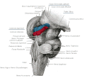
 Brainstem subregions of a healthy participant.jpg 1,995 × 2,188; 191 KB
-
Brainstem-and-Spinal-Cord-Circuitry-Regulating-REM-Sleep-and-Muscle-Atonia-pone.0024998.s001.ogv 16 s, 1,664 × 860; 1.39 MB
-
 Brainstem.png 800 × 455; 326 KB
-
 Coupe vertico-médiane ou sagittale.png 847 × 642; 921 KB
-
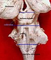
 Duret.0655.jpg 893 × 683; 94 KB
-
 EB1911 Brain Fig. 2-Medulla, Pons.jpg 525 × 442; 51 KB
-
 Enbor Entzefalikoa Atze ikuspegia.png 266 × 507; 54 KB
-
 Enbor Entzefalikoa Aurre ikuspegia.png 182 × 404; 43 KB
-
 Entzefalo enborra.png 415 × 324; 91 KB
-
 Entzefalo-enborra.png 391 × 321; 107 KB
-
-
 Ex vivo Brainstem sample.jpg 579 × 558; 259 KB
-
 External face of brainstem.jpg 1,700 × 2,338; 1.65 MB
-
 External view of basal ganglia.jpg 1,700 × 2,338; 1.62 MB
-
 Face antérieure du tronc cérébral.jpg 1,700 × 2,338; 1.69 MB
-
 Face latérale du tronc cérébral.jpg 1,700 × 2,338; 1.41 MB
-
 Face postérieure du tronc cérébral.jpg 1,700 × 2,338; 1.8 MB
-
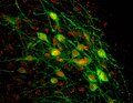
 FoxP2+TH sagittal.jpg 1,973 × 1,614; 485 KB
-
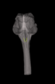
 GarpenBrain.jpg 1,280 × 1,024; 677 KB
-
 Gray681.png 600 × 527; 51 KB
-
 Gray682.png 413 × 800; 82 KB
-
 Gray684.png 533 × 800; 97 KB
-
 Gray685.png 453 × 750; 75 KB
-
 Gray691.png 500 × 570; 64 KB
-
 Gray692.png 175 × 500; 17 KB
-
 Gray697.png 450 × 486; 18 KB
-
 Gray705.png 500 × 374; 42 KB
-
 Gray719.png 550 × 503; 54 KB
-
 Green-HSD2 red-MR in NTS.jpg 631 × 491; 35 KB
-
 Hierarchy animate.gif 600 × 184; 630 KB
-
 Human brain left midsagitttal view closeup description 2.JPG 701 × 490; 61 KB
-
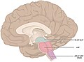
 Human brain left midsagitttal view closeup.JPG 701 × 490; 53 KB
-
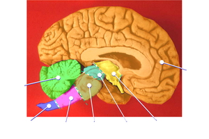
 Human brainstem Sagittal view.jpg 490 × 360; 146 KB
-
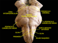
 Human brainstem-thalamus posterior view description.JPG 340 × 485; 23 KB
-
 Image from page 632 of "Human physiology" (1856) (14762204386).jpg 1,518 × 1,598; 285 KB
-
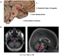
 Journal.pone-IT.png 1,044 × 958; 545 KB
-
 Journal.pone.0008247.g001.png 1,958 × 1,868; 1.19 MB
-
 Nerbio bagoaren bizkarraldeko nukleoa.png 340 × 524; 54 KB
-
 Nukleo anbiguoa bakarrik.png 261 × 422; 48 KB
-
 Nukleo anbiguoa, Alboko ikuspgia.png 473 × 577; 72 KB
-
 Nukleo anbiguoa, Aurreko ikuspegia.png 479 × 579; 222 KB
-
 Núcleos del rafé.png 1,104 × 907; 164 KB
-
 Physiology and biochemistry in modern medicine (1918) (14781026312).jpg 682 × 1,616; 119 KB
-
 Sistema nerviós central i parts del tronc encefàlic.png 401 × 374; 62 KB
-
 Slide2RAFA-es.png 1,000 × 750; 768 KB
-
 Slide2RAFA.JPG 960 × 720; 61 KB
-
 Sobo 1909 648.png 1,063 × 1,048; 3.19 MB
-
 Sobo 1909 660.png 864 × 890; 2.2 MB
-
 Sobo 1909 661.png 676 × 889; 1.72 MB
-
 Sobo 1909 676.png 1,944 × 1,764; 9.83 MB
-
 Solco bulbo-pontino.jpg 1,280 × 720; 117 KB
-
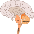
 Subcortical structures.png 646 × 200; 129 KB
-
 Substantia nigra pars compacta.jpg 451 × 377; 36 KB
-
 Tapatu gabe.tif 1,286 × 872; 210 KB
-
 The chemical neuroanatomy of the brainstem reticular formation (BRF).png 1,958 × 966; 806 KB
-

-

-
 Trigeminoa.jpg 657 × 621; 24 KB
-
 Tronc cereb lateral.jpg 316 × 269; 11 KB
-
 Tronc cerebral.JPG 338 × 397; 25 KB
-
 Tronc-cerebral-ventrale.jpg 370 × 410; 21 KB
-
 Tronco encefalico.gif 550 × 503; 54 KB


_(14581496279).jpg)


_(14762204386).jpg)


_(14781026312).jpg)


.png)
_(14750147536).jpg)
_(14770003121).jpg)


