Category:Illustrations by Drpaulineneveu
Media in category "Illustrations by Drpaulineneveu"
The following 83 files are in this category, out of 83 total.
-
 DrPaulineNeveu 01 Champs recepteurs simple complexe Receptor field.svg 1,002 × 844; 38 KB
-
 DrPaulineNeveu 01 Frequence potentiel action potential frequency.svg 1,294 × 1,051; 37 KB
-
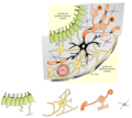 DrPaulineNeveu 01 GlieSNC cellules separees.png 732 × 653; 206 KB
-
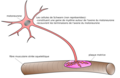
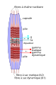
 DrPaulineNeveu 01 MyeliniqSchwann.png 630 × 501; 71 KB
-
 DrPaulineNeveu 01 NeuroneBipolaire.svg 827 × 397; 44 KB
-
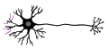
 DrPaulineNeveu 01 NeuroneMultipolaire.svg 772 × 703; 45 KB
-
 DrPaulineNeveu 01 NeuronePseudoUnipolaire.svg 716 × 260; 9 KB
-
 DrPaulineNeveu 01 Rubber hand illusion main caoutchouc.svg 443 × 417; 24 KB
-
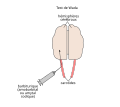
 DrPaulineNeveu 02 Iris sphincter dilator.svg 1,455 × 1,001; 14 KB
-
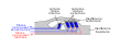
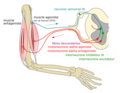
 DrPaulineNeveu 02 Jonction neuromusculaire.png 1,013 × 655; 149 KB
-
 DrPaulineNeveu 02 nobel.png 533 × 515; 66 KB
-
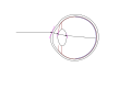
 DrPaulineNeveu 02 Oeil coupe.svg 1,075 × 591; 28 KB
-
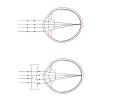
 DrPaulineNeveu 02 Oeil eye 3 plans refraction.svg 563 × 384; 6 KB
-
-

-
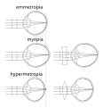
 DrPaulineNeveu 02 Oeil eye emmetrope.svg 653 × 390; 9 KB
-
 DrPaulineNeveu 02 Oeil eye hypermetrope hypermetropia.svg 888 × 634; 16 KB
-
 DrPaulineNeveu 02 Oeil eye myope myopia.svg 678 × 561; 16 KB
-
 DrPaulineNeveu 02 Oeil eye vision pres loin presbytie presbytia.svg 1,035 × 601; 16 KB
-
 DrPaulineNeveu 02 PA trace.svg 899 × 1,505; 108 KB
-
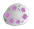
 DrPaulineNeveu 02 Recepteurs senso nue encaps cell.png 960 × 812; 143 KB
-
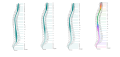 DrPaulineNeveu 03 Allometrie ME et colonne vertebrale.svg 1,872 × 882; 375 KB
-
 DrPaulineNeveu 03 Apparition Vesicules.png 1,258 × 623; 199 KB
-
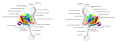
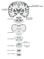
 DrPaulineNeveu 03 Axe hypothalamus hypophyse.png 1,078 × 848; 306 KB
-
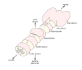
 DrPaulineNeveu 03 Cellules ciliees int ext et mb tectoriale.svg 589 × 218; 29 KB
-
 DrPaulineNeveu 03 EEG potentiels evoques.svg 683 × 369; 66 KB
-
 DrPaulineNeveu 03 Embryo SNC courbures.svg 516 × 865; 29 KB
-
 DrPaulineNeveu 03 Humain profil.png 244 × 841; 50 KB
-
 DrPaulineNeveu 03 Hypothalamus epaisseur noyaux.svg 1,723 × 628; 140 KB
-
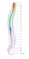 DrPaulineNeveu 03 Myelomeres et dermatomes.svg 404 × 825; 139 KB
-
 DrPaulineNeveu 03 SNC LCR circulation.svg 200 × 387; 37 KB
-
 DrPaulineNeveu 03 Tube neural cadre d etude.svg 647 × 550; 44 KB
-
 DrPaulineNeveu 03 Voies nociceptives pathways tracks.svg 585 × 579; 842 KB
-
 DrPaulineNeveu 04 Langue et gouts.svg 277 × 618; 24 KB
-
 DrPaulineNeveu 05 Actine molecule.svg 813 × 157; 29 KB
-
 DrPaulineNeveu 05 Contraction muscle cinq etapes.svg 3,597 × 1,149; 97 KB
-
 DrPaulineNeveu 05 Fibre musculaire entiere schematisee.svg 255 × 838; 17 KB
-
 DrPaulineNeveu 05 myosine bouquet.svg 477 × 168; 11 KB
-
 DrPaulineNeveu 05 Myosine molecule.svg 520 × 223; 14 KB
-
 DrPaulineNeveu 05 Sarcomere schema actine myosine.svg 547 × 327; 239 KB
-
 DrPaulineNeveu 05 Secousse fibres rapide interm lente.svg 945 × 832; 143 KB
-
 DrPaulineNeveu 05 Secousse musculaire.svg 575 × 249; 14 KB
-
 DrPaulineNeveu 05 Secousse sommation.svg 500 × 638; 24 KB
-
 DrPaulineNeveu 05 Tetanos.svg 905 × 361; 35 KB
-
 DrPaulineNeveu 05 Tissu conjonctif endo peri epimysium.svg 593 × 602; 408 KB
-
 DrPaulineNeveu 06 Homonculus senso et moteur.svg 563 × 282; 54 KB
-
 DrPaulineNeveu 06 UM.svg 854 × 421; 27 KB
-
 DrPaulineNeveu 06 Voies lemniscales.svg 367 × 533; 847 KB
-
 DrPaulineNeveu 06 Voies spino cerebelleuses.svg 343 × 521; 835 KB
-
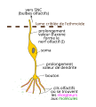
 DrPaulineNeveu 07 FNM detail.svg 611 × 921; 96 KB
-
 DrPaulineNeveu 07 Locomotion mvt corps.svg 375 × 248; 9 KB
-
 DrPaulineNeveu 07 Marche humaine poser lever.svg 569 × 209; 58 KB
-
 DrPaulineNeveu 07 Mb sup Reflexe Inhibition autogeniq.png 1,044 × 810; 215 KB
-
 DrPaulineNeveu 07 Mb sup Reflexe Myotatique.png 1,098 × 818; 221 KB
-
 DrPaulineNeveu 07 Neurone olfactif.svg 486 × 542; 78 KB
-
 DrPaulineNeveu 07 OTG detail.png 409 × 626; 85 KB
-
 DrPaulineNeveu 07 Poser lever pendule pendule inverse.svg 230 × 162; 10 KB
-
 DrPaulineNeveu 07 Voies gustatives pathways tracks.svg 381 × 511; 813 KB
-
 DrPaulineNeveu 08 Ampoule canal.png 587 × 797; 87 KB
-
 DrPaulineNeveu 08 Cellule ciliee cupule.png 399 × 860; 151 KB
-
 DrPaulineNeveu 08 Inclinaison cils.svg 685 × 506; 115 KB
-
 DrPaulineNeveu 08 Inclinaison tete et acceleration voiture.png 1,055 × 739; 281 KB
-
 DrPaulineNeveu 08 Liens Vestib Muscles oculomoteurs.svg 309 × 339; 111 KB
-
 DrPaulineNeveu 08 Voies Vestibulaires.svg 324 × 512; 832 KB
-
 DrPaulineNeveu 09 Voies Reticulaires.svg 315 × 493; 819 KB
-
 DrPaulineNeveu 10 Voies CorticoNucleaires.svg 585 × 579; 820 KB
-
 DrPaulineNeveu 10 Voies CorticoReticuloSpinales.svg 585 × 579; 816 KB
-
 DrPaulineNeveu 10 Voies CorticoRubroSpinales.svg 585 × 579; 812 KB
-
 DrPaulineNeveu 10 Voies CorticoSpinales.svg 585 × 579; 830 KB
-
 DrPaulineNeveu 10 Voies CorticoTectoSpinales.svg 585 × 579; 816 KB
-
 DrPaulineNeveu 10 Voies CorticoVestibuloSpinales.svg 585 × 579; 814 KB
-
 DrPaulineNeveu 12 OldsMilnerSelfStimulation.svg 927 × 639; 36 KB
-
 DrPaulineNeveu 13 Voies dorsale ventrale.svg 822 × 543; 19 KB
-
 DrPaulineNeveu 14 EEG technique.svg 551 × 265; 56 KB
-
 DrPaulineNeveu 14 Modes transmission burst.svg 550 × 813; 41 KB
-
 DrPaulineNeveu 15 PLT.svg 551 × 774; 72 KB
-
 DrPaulineNeveu 15 Synapse CA3 CA1.svg 703 × 320; 18 KB
-
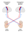 DrPaulineNeveu 16 Controle controlateral mains.svg 526 × 646; 61 KB
-
 DrPaulineNeveu 16 Hemichamps visuels point fixation.svg 510 × 438; 24 KB
-
 DrPaulineNeveu 16 Test Wada.svg 437 × 389; 17 KB
-
 DrPaulineNeveu Schwann cell cellule unmyelinated amyelinique.svg 412 × 346; 68 KB
-
 DrPaulineNeveuNeuroneEpineDendritique.svg 716 × 324; 30 KB
-
 DrPaulineNeveuNeuroneMultipolaireGaineMyéline.svg 677 × 314; 25 KB


