Category:Medical illustrations of tongues
Media in category "Medical illustrations of tongues"
The following 124 files are in this category, out of 124 total.
-
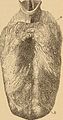
 Anatomical model of human tongue Wellcome L0010002.jpg 1,598 × 1,182; 408 KB
-
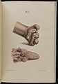
 Anatomical model of human tongue Wellcome L0010003.jpg 1,098 × 1,688; 415 KB
-
 Anatomy, physiology and hygiene for high schools (1900) (14594907458).jpg 868 × 1,648; 551 KB
-
 Anesthetic and tubercular leprosy Wellcome L0074854.jpg 5,336 × 7,639; 6.63 MB
-
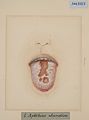 Aphthous ulceration of the tongue Wellcome L0062793.jpg 3,723 × 5,023; 2.3 MB
-
 Bald areas on tongue Wellcome L0061273.jpg 4,008 × 4,908; 3.16 MB
-
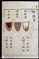 C14 Chinese tongue diagnosis chart Wellcome L0039594.jpg 2,083 × 3,125; 5.64 MB
-
 C14 Chinese tongue diagnosis chart Wellcome L0039595.jpg 2,083 × 3,125; 5.43 MB
-
 C14 Chinese tongue diagnosis chart Wellcome L0039596.jpg 2,083 × 3,125; 5.55 MB
-
 C14 Chinese tongue diagnosis chart Wellcome L0039597.jpg 2,083 × 3,125; 4.79 MB
-
 C14 Chinese tongue diagnosis chart Wellcome L0039598.jpg 2,083 × 3,125; 5.05 MB
-
 C18 Chinese woodcut; Laryngitis extending to the tongue Wellcome L0039732.jpg 2,098 × 3,146; 5.27 MB
-
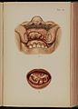
 Cancer of the tongue Wellcome L0061564.jpg 4,290 × 6,186; 3.5 MB
-
 Chinese C19 woodcut; Abscesses beneath the tongue Wellcome L0038870.jpg 2,069 × 3,104; 2.75 MB
-
 Chinese tongue diagnosis diagram Wellcome L0039592.jpg 2,083 × 3,125; 5.51 MB
-
 Chinese tongue diagnosis diagram; 'Baked centre' tongue' Wellcome L0039589.jpg 2,083 × 3,125; 4.98 MB
-
 Chinese tongue diagnosis diagram; 'Black within' tongue Wellcome L0039593.jpg 2,083 × 3,125; 5.16 MB
-
 Chinese tongue diagnosis diagram; 'Incipient plague' tongue Wellcome L0039588.jpg 2,083 × 3,125; 5.26 MB
-
 Chinese tongue diagnosis diagram; 'Red star' tongue Wellcome L0039591.jpg 2,083 × 3,125; 5.07 MB
-
 Chinese tongue diagnosis diagram; 'Spotted tongue' Wellcome L0039590.jpg 2,083 × 3,125; 5.25 MB
-
 Chinese tongue diagnosis diagram; 'White-coated tongue' Wellcome L0039587.jpg 2,083 × 3,125; 5.21 MB
-
 Congenital pedunculated tumour of the tongue Wellcome L0062772.jpg 4,324 × 5,884; 3.42 MB
-
 Cyst under the tongue of an infant Wellcome L0061280.jpg 3,589 × 5,297; 3.99 MB
-
 Degenerated naevus on the tongue Wellcome L0061284.jpg 3,978 × 4,926; 3.9 MB
-
 Dental ulcer on the tongue Wellcome L0062797.jpg 4,035 × 5,059; 3.16 MB
-
 Different stages of yellow fever, 1820 Wellcome L0074834.jpg 5,711 × 7,410; 6.76 MB
-
 Double page spread from Japanese MS 76 Wellcome L0031458.jpg 3,779 × 2,981; 4.41 MB
-
 Dyspeptic ulcers of the tongue Wellcome L0062738.jpg 3,964 × 5,516; 4.23 MB
-
 Effects of tertiary syphilis of the tongue Wellcome L0062792.jpg 3,601 × 5,398; 2.85 MB
-
 Epithelioma and ichthyosis of the tongue Wellcome L0061288.jpg 3,757 × 4,877; 3.6 MB
-
 Excoriated tongue Wellcome L0062788.jpg 3,504 × 5,256; 2.94 MB
-
 Extensive epithelioma of the dorsum of the tongue Wellcome L0062782.jpg 4,685 × 3,960; 3.84 MB
-
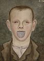 Face of a boy suffering from purpura haemorrhagica Wellcome L0062019.jpg 4,184 × 5,836; 6.35 MB
-
 Gangrenous tonsils Wellcome L0061523.jpg 4,244 × 5,664; 3.64 MB
-
 Gummatous ulcer of the tongue Wellcome L0062802.jpg 4,279 × 5,673; 3.45 MB
-
 Gummatous ulcer of the tongue Wellcome L0062803.jpg 4,217 × 6,141; 3.75 MB
-
 Gummatous ulceration of the tongue Wellcome L0061561.jpg 4,733 × 6,210; 3.48 MB
-
 Gums and tongue from a case of lead poisoning Wellcome L0062323.jpg 3,844 × 5,152; 4.77 MB
-
 Hemiatrophy and hemiplegia of the left side of the tongue Wellcome L0062734.jpg 4,997 × 4,363; 3.69 MB
-
 Hunterian chancre on the tongue Wellcome L0062950.jpg 3,796 × 4,737; 2.41 MB
-
 Ichthyosis of the tongue Wellcome L0062806.jpg 4,129 × 5,309; 3.27 MB
-
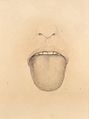
 Indented tongue Wellcome L0062787.jpg 3,757 × 5,197; 2.14 MB
-
 Inflamed dental ulcer on the tongue Wellcome L0062798.jpg 4,311 × 5,507; 3.72 MB
-
 Inflammatory hypertrophy of the tongue Wellcome L0061277.jpg 4,014 × 4,848; 4.15 MB
-
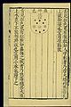
 Japanese MS 94 Wellcome L0026717.jpg 1,580 × 1,238; 580 KB
-
 Kawasaki symptoms B.jpg 464 × 384; 16 KB
-
 Large, soft naevus on the tongue Wellcome L0061282.jpg 3,923 × 4,787; 3.33 MB
-
 Leucoma and epithelioma of the tongue Wellcome L0061266.jpg 4,044 × 4,896; 4.12 MB
-
 Leucoma and inflammation of the tongue Wellcome L0062808.jpg 4,295 × 6,089; 4.31 MB
-
 Leucoma on the tongue with fluffy surface Wellcome L0062805.jpg 4,269 × 5,673; 3.64 MB
-
 Lupus ulcer on the tongue Wellcome L0062800.jpg 4,529 × 5,944; 3.78 MB
-
 Lymphangioma occupying the right side of the tongue Wellcome L0062764.jpg 3,893 × 5,309; 2.78 MB
-
 Lymphangioma of the left side of the tongue Wellcome L0062760.jpg 4,077 × 4,326; 2.85 MB
-
 Lymphangioma of the tongue Wellcome L0062761.jpg 3,209 × 3,617; 1.33 MB
-
 Lymphangioma of the tongue Wellcome L0062762.jpg 2,833 × 2,324; 1.11 MB
-
 Lymphangioma on the tip of the tongue Wellcome L0062766.jpg 4,616 × 4,724; 2.94 MB
-
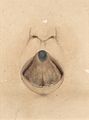
 Mouth with large sublingual cyst Wellcome L0062758.jpg 4,248 × 4,342; 3.52 MB
-
 Naevus on the tip of the tongue of an old woman Wellcome L0061281.jpg 3,718 × 4,998; 3.16 MB
-
 Naevus on the tongue Wellcome L0061283.jpg 3,678 × 6,324; 3.85 MB
-
 Nigrities linguae Wellcome L0062742.jpg 3,664 × 5,596; 4.49 MB
-
 Nigrities linguae Wellcome L0062743.jpg 4,412 × 4,616; 3.43 MB
-
 Page from Japanese MS 76 Wellcome L0031457.jpg 2,682 × 4,056; 4.38 MB
-
 Plate VIII, mouth & face lesions Prince Albert Morrow, 1889 Wellcome L0074356.jpg 5,181 × 7,053; 9.23 MB
-
 Radium, 1917; photos of tongue cancer Wellcome L0016772.jpg 1,702 × 1,132; 1.01 MB
-
 Ranula under the tongue of a young man Wellcome L0061279.jpg 3,619 × 5,725; 4.59 MB
-
 Rapidly growing epithelioma of the tongue Wellcome L0061563.jpg 4,734 × 6,102; 4.29 MB
-
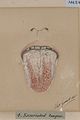
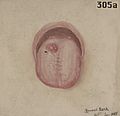
 Scorbutic tongue (cropped).jpg 209 × 195; 8 KB
-
 Scorbutic tongue.jpg 700 × 468; 56 KB
-
 Small lymphangioma of the tongue Wellcome L0062765.jpg 4,266 × 5,238; 2.7 MB
-
 Smooth tongue from an anaemic woman Wellcome L0061274.jpg 3,859 × 5,171; 2.57 MB
-
 Squamous celled carcinoma of the tongue Wellcome L0061285.jpg 3,876 × 4,758; 4.03 MB
-
 Squamous celled carcinoma of the tongue Wellcome L0061287.jpg 4,102 × 5,081; 3.62 MB
-
 Stomach, with mucous membrane thickened and mammillated Wellcome L0061177.jpg 4,172 × 4,884; 4.42 MB
-
 Syphilis; lesion on man's tongue, 1858 Wellcome V0010159.jpg 3,152 × 2,876; 3.48 MB
-
 Syphilis; lesions on tongue and upper lip, 1869 Wellcome V0010160.jpg 2,610 × 3,516; 3.82 MB
-
 Syphilis; lesions on tongue, 1857 Wellcome V0010154.jpg 2,515 × 2,795; 2.79 MB
-
 Syphilis; lesions on woman's tongue, 1866 Wellcome V0010158.jpg 2,250 × 3,123; 2.68 MB
-
 Syphilis; lesions under tongue, 1856 Wellcome V0010161.jpg 2,481 × 3,474; 3.74 MB
-
 Tab 10, Sclerosis of tongue, Mracek, 1898 Wellcome L0074268.jpg 8,016 × 5,876; 16.11 MB
-
 Tab 41, Infiltration, submucosa, mouth. Mracek 1898 Wellcome L0074258.jpg 5,702 × 7,950; 12.87 MB
-
 Tertiary syphilitic disease of the tongue Wellcome L0062750.jpg 4,488 × 4,140; 3.63 MB
-
 Tertiary syphilitic disease of the tongue Wellcome L0062752.jpg 4,780 × 4,272; 3.59 MB
-
 Tertiary syphilitic disease of the tongue Wellcome L0062753.jpg 3,640 × 4,893; 3.52 MB
-
 Tertiary syphilitic disease of the tongue Wellcome L0062754.jpg 4,482 × 4,233; 3.22 MB
-
 Tertiary syphilitic disease of the tongue Wellcome L0062755.jpg 5,020 × 4,083; 3.72 MB
-
 Tertiary syphilitic ulceration of the tongue Wellcome L0062804.jpg 4,669 × 6,401; 3.42 MB
-
 The diseased tissue around the anus and genitals of a woman, Wellcome V0009921.jpg 3,060 × 2,432; 3.54 MB
-
 The lower half of a face with a skin disease on the top lip; Wellcome V0010255.jpg 2,328 × 3,250; 2.66 MB
-
 The upper half of a face with a skin disease on the forehead Wellcome V0010258.jpg 3,250 × 2,307; 2.79 MB
-
 Tongue and fauces from a case of diphtheria Wellcome L0062739.jpg 4,072 × 5,852; 3.51 MB
-
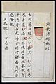
 Tongue diagnosis chart, Chinese woodcut, late Ming Wellcome L0038015.jpg 1,984 × 2,976; 2.24 MB
-
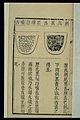
 Tongue diagnosis chart, Chinese woodut, late Ming Wellcome L0038014.jpg 1,984 × 2,976; 2.56 MB
-
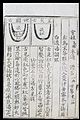
 Tongue diagnosis chart; Long, swollen, red, protruding tongue Wellcome L0038681.jpg 2,069 × 3,104; 2.67 MB
-
 Tongue diagnosis chart; Red; white coating; stringy mucus Wellcome L0038682.jpg 2,069 × 3,104; 2.82 MB
-
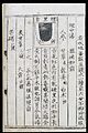
 Tongue morphology, Chinese woodcut Wellcome L0038024.jpg 1,984 × 2,976; 2.8 MB
-
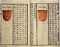
 Tongue morphology; Cidi heishe) and heilan zi nie she Wellcome L0038023.jpg 1,984 × 2,976; 2.59 MB
-
 Tongue morphology; Heitai biandi hongshe, Chinese woodcut Wellcome L0038022.jpg 1,984 × 2,976; 2.71 MB
-
 Tongue of a boy in a state of acute glossitis Wellcome L0062735.jpg 5,091 × 4,222; 4.13 MB
-
 Tongue of a boy with 'wandering rash' Wellcome L0061268.jpg 3,705 × 5,190; 3.05 MB
-
 Tongue of a child with Bright's disease Wellcome L0062736.jpg 4,763 × 4,300; 3.41 MB
-
 Tongue of a man with a tubercular ulcer Wellcome L0062745.jpg 4,579 × 4,445; 3.87 MB
-
 Tongue showing an ichthyotic condition Wellcome L0062778.jpg 4,827 × 4,303; 4.38 MB
-
 Tongue with a leucomatous condition of the dorsum Wellcome L0062779.jpg 4,560 × 4,540; 2.92 MB
-
 Tongue with a primary syphilitic sore Wellcome L0062749.jpg 4,096 × 4,832; 3.04 MB
-
 Tongue with a recurrent papillary growth Wellcome L0062767.jpg 4,004 × 5,824; 3.21 MB
-
 Tongue with a recurrent papillary growth Wellcome L0062768.jpg 4,052 × 5,756; 2.95 MB
-
 Tongue with bluish naevoid tumour Wellcome L0062771.jpg 4,670 × 4,245; 2.94 MB
-
 Tongue with chronic ulcer Wellcome L0062795.jpg 3,629 × 5,021; 1.96 MB
-
 Tongue with diphtheritic membrane Wellcome L0062740.jpg 3,824 × 5,564; 2.98 MB
-
 Tongue with dyspeptic ulcer Wellcome L0062796.jpg 4,529 × 6,204; 3.75 MB
-
 Tongue with furrows of tertiary syphilis Wellcome L0062791.jpg 3,601 × 5,398; 2.59 MB
-
 Tongue with primary syphilitic sore on the tongue Wellcome L0062951.jpg 4,602 × 4,409; 2.3 MB
-
 Tongue with small multiple ulcers on the dorsum linguae Wellcome L0062737.jpg 4,469 × 4,550; 4.12 MB
-
 Tongue with tertiary syphilitic ulcer Wellcome L0062751.jpg 3,724 × 5,832; 3.19 MB
-
 Tongue with tuberculous ulcer Wellcome L0062799.jpg 4,243 × 6,245; 3.37 MB
-
 Tongue with two large ulcers due to congenital syphilis Wellcome L0062747.jpg 4,184 × 4,787; 3.97 MB
-
 Tongue, mouth, and moustache of a man with skin disease Wellcome L0060799.jpg 6,584 × 4,157; 4.68 MB
-
 Tubercular ulceration of the dorsum of the tongue Wellcome L0062744.jpg 3,804 × 5,584; 3.54 MB
-
 Ulcerated mucous patches on the tongue Wellcome L0062801.jpg 4,503 × 5,684; 4.07 MB
-
 Unilateral atrophy of the tongue Wellcome L0061560.jpg 3,832 × 5,394; 3.44 MB
-
 Vesicles on the tongue preceding aphthae Wellcome L0062786.jpg 3,769 × 5,241; 2.21 MB
-
 Warty, papillated growth on tongue Wellcome L0062780.jpg 3,492 × 5,792; 2.98 MB
-
 Woman with a medullary cancerous growth on the tongue Wellcome L0061161.jpg 4,622 × 5,453; 3.55 MB
-
 Xanthelasma of the tongue Wellcome L0062741.jpg 6,040 × 4,204; 4.15 MB


_(14594907458).jpg)


.jpg)


_and_heilan_zi_nie_she_Wellcome_L0038023.jpg)


