Category:Mitotic cells stained with DAPI
Media in category "Mitotic cells stained with DAPI"
The following 25 files are in this category, out of 25 total.
-
 3D-SIM-4 Anaphase 3 color.jpg 954 × 724; 264 KB
-
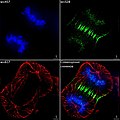
 Binucleated cell overlay.tiff 1,392 × 1,040; 4.17 MB
-
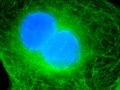
 Binucleated cell.jpg 514 × 386; 45 KB
-
 Cell divisions in Arabidopsis primary root meristem cells.tif 1,527 × 513; 765 KB
-
 Chromatin bridge stained with DAPI 2.tiff 1,392 × 1,040; 4.14 MB
-
 Cortical and mitotic microtubules in Arabidopsis primary root meristem cells.tif 1,525 × 504; 1,003 KB
-
 Cutie in mitosis.jpg 1,004 × 1,002; 835 KB
-
 Different mitotic stages in Arabidopsis primary root meristem cells.tif 774 × 256; 287 KB
-
 Dividing Cell Fluorescence-ru.jpg 554 × 554; 165 KB
-
 Dividing Cell Fluorescence-uk.jpg 554 × 554; 184 KB
-
 Dividing Cell Fluorescence.jpg 554 × 554; 58 KB
-
-
 Endogenous hMad1.png 575 × 873; 511 KB
-

-

-
 MAX MI DAPI 9-07-2015 A2 well.png 9,136 × 9,116; 62.29 MB
-
 Metaphase anaphase.png 232 × 153; 28 KB
-
 Mitosepanel es.jpg 1,050 × 246; 27 KB
-
 Mitosepanel-rus.tif 941 × 234; 234 KB
-
 Mitosepanel.jpg 1,050 × 246; 39 KB
-

-
 Mitotic spindle in Arabidopsis primary root meristem cells.tif 615 × 201; 195 KB
-
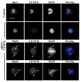 SiCENP-E metaphase.png 501 × 510; 97 KB
-
 Spindle with Four Poles (8744518858).jpg 448 × 446; 93 KB
-
 The Biological bulletin (20353224406).jpg 1,806 × 1,582; 1,022 KB


_and_chromosomes_(red-yellow).png)


.jpg)
.jpg)