Category:National finalists for the Microscopy category from Wiki Science Competition 2023
Media in category "National finalists for the Microscopy category from Wiki Science Competition 2023"
The following 49 files are in this category, out of 49 total.
-
 . Bothriocephalus opsariichthydis - whole parasite (27 x).tif 1,021 × 723; 723 KB
-
 Acido acetilsalicilico.jpg 3,959 × 2,640; 2.21 MB
-
 Ag micromirrors BSE.jpg 2,048 × 2,048; 2.09 MB
-
 Ammophila arenaria transverse section.jpg 3,622 × 3,598; 9.35 MB
-
 Amyloid deposition in prostate tumor tissue.jpg 4,096 × 3,008; 2.15 MB
-
 Apareamiento de copépodos Boeckella gracilis.jpg 7,952 × 6,874; 9.42 MB
-
 Approaching.png 1,280 × 955; 2.59 MB
-
 Argulus foliaceus (Linnaeus, 1758) - abdomen.tif 1,019 × 721; 718 KB
-
 AZM Euplotes vanleeuwenhoeki.tif 1,494 × 1,996; 2.02 MB
-
 Bond Forever.png 1,005 × 630; 1.2 MB
-
 Bryophyte Leaf Cells.jpg 2,982 × 1,870; 4.94 MB
-
 Ceriodaphnia campo oscuro.jpg 2,492 × 2,805; 1.48 MB
-
 Colloid lithography, artificial opal, polystyrene.jpg 2,880 × 2,160; 4.99 MB
-
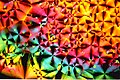
 Cristales de ácido ascórbico (Vitamina C) vistos con lu polarizada.jpg 1,800 × 4,000; 3.78 MB
-
 Cymbella peraspera.jpg 3,008 × 3,000; 1.03 MB
-
 Daphnia con huevos en campo oscuro.jpg 6,000 × 6,000; 6.79 MB
-
 Degenerating-drosophila-retina.jpg 10,560 × 10,672; 76.6 MB
-
 Differentiation of Human-Induced Pluripotent Stem Cells to GABAergic Neurons.tif 1,296 × 966; 3.58 MB
-
 Dorsal part of Argulus foliaceus.tif 1,021 × 723; 723 KB
-
 Embryo in flower.png 3,000 × 3,006; 2.97 MB
-
 Fibrin clot formed in the presence of F(ab) fragments of monoclonal antibody III-1D.jpg 1,696 × 2,090; 723 KB
-
 Fluorescence.microscope3.jpg 2,436 × 2,393; 1.26 MB
-
 Iron Sulphate Nucleation Point.jpg 2,982 × 1,989; 5.16 MB
-
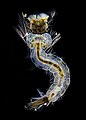 Larva de mosquito campo osuro.jpg 9,694 × 13,500; 32.42 MB
-
 Lead Trees in a Displacement Reaction.jpg 6,000 × 4,000; 8.78 MB
-
 Metaphase meiosis II.png 1,392 × 1,024; 1.15 MB
-
 Micro-Girasoli.jpg 2,355 × 2,355; 4.45 MB
-
 Mortal dance - The Sinister Beauty of the Infection.jpg 1,300 × 1,150; 1.2 MB
-
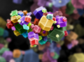 Nano-Legos.png 3,000 × 2,236; 4.34 MB
-
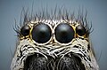 Oczy pająka z rodziny skakunowatych.jpg 3,500 × 2,308; 5 MB
-
 Oxidized surface of copper.jpg 6,000 × 4,000; 12.11 MB
-
 Parasites in freshwater mussel 03.jpg 1,080 × 810; 101 KB
-
 Pistesääsklase Culiceta annulata tiib.jpg 6,000 × 2,526; 13.95 MB
-
 Pitting on copper - Piqûre de corrosion sur du cuivre.jpg 2,560 × 1,920; 1.48 MB
-
 Polilla Mythimna loreyi microscopio electrónico de barrido.jpg 3,072 × 2,095; 7.66 MB
-
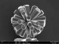
 Pupae of the parasitoid Paracodrus apterogynus.jpg 4,658 × 2,620; 1.74 MB
-
 Pyramids of anhydrite.jpg 4,908 × 3,264; 12.29 MB
-
 Płatek sniegu.jpg 3,500 × 2,333; 5.23 MB
-
 Science & Nature.jpg 2,048 × 2,048; 2.48 MB
-
 SEM Zircon separation.jpg 2,090 × 2,785; 1.29 MB
-
 SiC Pumpkin.png 1,280 × 940; 1.71 MB
-
 Spinal cord gray matter immunofluorescence staining, confocal imaging.png 1,600 × 1,600; 5.88 MB
-
 Tiigi klaasiksääsevastne.jpg 4,763 × 3,176; 7.04 MB
-
 Vanadium crystal.jpg 5,021 × 2,000; 1.54 MB
-
 Vitamina C.jpg 3,502 × 2,334; 1.24 MB
-
 ZnOGO-031.tif 1,924 × 1,443; 2.56 MB
-
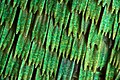 Łuski na skrzydle motyla.jpg 3,500 × 2,340; 8.17 MB
-
 Кромид под микроскоп.jpg 1,600 × 1,200; 140 KB
-
 Лист од лопен- verbascum sp.jpg 1,600 × 1,200; 135 KB


_vistos_con_lu_polarizada.jpg)


_fragments_of_monoclonal_antibody_III-1D.jpg)


