Category:Neuroimaging
set of techniques to measure and visualize aspects of the nervous system | |||||
| Upload media | |||||
| Instance of | |||||
|---|---|---|---|---|---|
| Subclass of | |||||
| |||||
Subcategories
This category has the following 22 subcategories, out of 22 total.
*
B
D
E
F
- FMRI BOLD (6 F)
K
M
- Media from NeuroImage (6 F)
N
O
- Optical imaging (86 F)
S
- SQUID (27 F)
V
- Voxel-based morphometry (13 F)
Media in category "Neuroimaging"
The following 65 files are in this category, out of 65 total.
-
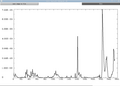
 Analyse spectrale d'un EEG.jpg 1,156 × 1,772; 361 KB
-

-
 Beta-CIT.png 808 × 484; 10 KB
-
 Blonde fNIRS lady.jpg 1,500 × 1,000; 175 KB
-
 BrainMRI3planes.gif 326 × 83; 1.91 MB
-
 Carte EEG sommeil paradoxal.jpg 768 × 576; 76 KB
-
 Coreg.png 557 × 525; 52 KB
-
 DCM for ERP and CMC.svg 640 × 436; 51 KB
-
 DCM for fMRI.png 1,657 × 1,780; 178 KB
-
 DCM for fMRI.svg 497 × 534; 19 KB
-
 DTschematic.jpg 578 × 616; 44 KB
-
 Ellipsoid eigenvalues14142.jpg 560 × 420; 20 KB
-
 Ellipsoid2722.jpg 560 × 420; 21 KB
-
 Ellipsoid888.jpg 560 × 420; 24 KB
-
 EP2D BOLD resting-state Siemens scanner sequence printout.png 827 × 1,169; 196 KB
-
 ExampleDCMGraph.png 378 × 189; 3 KB
-
 F3.medium.gif 440 × 316; 34 KB
-
 FEAT.png 435 × 446; 30 KB
-
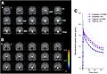
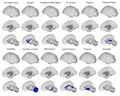
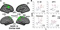 Fibromyalgia Glial Activation Agreement between SUVR and VT analyses A. Fig. 3.jpg 2,159 × 1,076; 237 KB
-

-
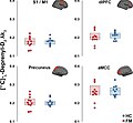
 Fibromyalgia Glial Activation Voxelwise group differences in (11C)PBR28 VT. A Fig. 1.jpg 2,130 × 1,104; 237 KB
-
-

-
 FNIRS head Hitachi ETG4000 2.jpg 3,264 × 1,836; 836 KB
-
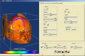
 FSL overview.png 719 × 625; 102 KB
-
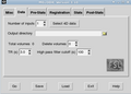
 FSL spatialsmoothing screenshot1.png 586 × 478; 83 KB
-
 FSL start GUI and motion correction buttion.png 663 × 491; 83 KB
-
 FSL toolbar.png 538 × 91; 20 KB
-
 Graph afni.png 699 × 499; 38 KB
-
 Haxby2001.jpg 763 × 500; 108 KB
-
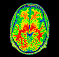
 Hirnperfusion normal.jpg 811 × 776; 261 KB
-
 Hirnperfusionsszintigrafie Hirntod Ausschnitt.jpg 387 × 747; 86 KB
-
 Hirnperfusionsszintigrafie Hirntod.jpg 825 × 777; 172 KB
-
 Hirnperfusionsszintigrafie normal Ausschnitt.jpg 386 × 745; 122 KB
-
 Invesalius.PNG 699 × 459; 214 KB
-
 Invesalius3 promed0446.png 1,280 × 779; 357 KB
-
 Kernel-logo.png 400 × 114; 13 KB
-
 LORETA qEEG in a person with ME-CFS small.gif 686 × 313; 97 KB
-
 LORETA qEEG in a person with ME-CFS.webp 1,292 × 588; 439 KB
-
 MELODIC.png 498 × 355; 27 KB
-
 Minc-n3-ex-corrected-spect.png 480 × 460; 217 KB
-
 Minc-n3-ex-corrected.png 480 × 460; 113 KB
-
 Minc-n3-ex-field.png 480 × 460; 59 KB
-
 Minc-n3-ex-original-spect.png 480 × 460; 219 KB
-
 Minc-n3-ex-original.png 480 × 460; 113 KB
-
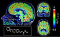
 MRI Brain Septum Deviation.png 766 × 947; 509 KB
-
 MRI EGC sagittal.png 576 × 576; 306 KB
-
 Mricron-slice-order-nifti.png 394 × 387; 33 KB
-
 MSTAT.jpg 668 × 416; 59 KB
-
 Neuroimaging Research Infographic.pdf 2,479 × 3,506, 2 pages; 1.21 MB
-
 Nirs.jpg 1,079 × 610; 312 KB
-
 Outlier afni.png 946 × 774; 14 KB
-
 Outlier despike.png 976 × 698; 14 KB
-
 PMC3492204 1750-1172-7-27-3.png 512 × 315; 155 KB
-
-
 Researcher-test.jpg 450 × 217; 50 KB
-
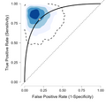 ROC.NateNet.png 530 × 530; 42 KB
-
 RTI-121.png 391 × 271; 5 KB
-
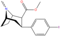
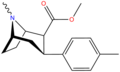
 RTI-229 structure.png 798 × 489; 11 KB
-
 RTI-32 structure.png 802 × 482; 10 KB
-
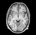
 Sagittal brain MRI.jpg 240 × 271; 15 KB
-
 Slicetime fsl.png 444 × 492; 31 KB
-

-
 Subcortical atlas regions.pdf 1,604 × 1,289; 4.61 MB
-
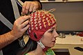 Unbonnetalectrodes.jpg 4,288 × 2,848; 4.97 MB


PBR28_SUVR_A._Fig._2.jpg)
PBR28_VT._A_Fig._1.jpg)


-PK11195_binding_in_CFS.ME_patient_(A)_and_healthy_control_(B).jpg)


-PK11195_in_CFS.ME_patients_and_healthy_controls.gif)
