Category:Petri dishes cultures with Antibiogram
Media in category "Petri dishes cultures with Antibiogram"
The following 63 files are in this category, out of 63 total.
-
 Ab Acinetobacter B 1.jpg 507 × 513; 79 KB
-
 Actionofgarlic1.jpg 2,448 × 3,264; 1.17 MB
-
 Anti1.tif 1,392 × 1,034; 1.38 MB
-
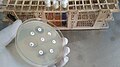
 Antibiogram Neomycin.jpg 682 × 682; 216 KB
-
 Antibiogram of Bacteria on MHA.jpg 4,000 × 2,250; 2.4 MB
-
 Antibiogram of Staphylococcus aureus on MHA.jpg 4,000 × 2,250; 2.33 MB
-
 Antibiogram of Viridans streptococci.jpg 4,000 × 3,000; 6.44 MB
-
 Antibiogram-Mueller-Hinton.JPG 3,648 × 2,736; 1.78 MB
-
 Antibiogramm Aspergillus-Pilz.jpg 1,936 × 2,592; 1.94 MB
-
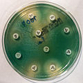
 Antibiogramma01.jpg 635 × 510; 248 KB
-
 Antibiograms.jpg 3,264 × 2,448; 1.2 MB
-
 Antibiotic disk diffusion.jpg 614 × 461; 142 KB
-
 Antibiotic resistant bacteria.jpg 1,280 × 1,024; 220 KB
-
 Antibiotic sensitivity and resistance.jpg 2,456 × 1,273; 870 KB
-
 Antibiotic suceptible bacteria.jpg 2,592 × 1,552; 2.42 MB
-
 Antibiotic susceptibility disk diffusion.jpg 2,592 × 1,552; 1.97 MB
-
 Antibiotics Sensitivity Testing (AST) Pattern of Enterococcus.jpg 4,000 × 2,250; 2.93 MB
-
 Antibiotics tested for beta-haemolytic streptococci.jpg 4,160 × 2,340; 3.13 MB
-
 AST on blood agar.jpg 2,592 × 1,552; 915 KB
-
 Bacillus growth inhibited by ciprofloxacin.jpg 448 × 336; 31 KB
-
 Bacterial lawn 01.jpg 2,992 × 2,012; 2.96 MB
-
 Bacteroides fragilis on Fastidious Anaerobe Agar - Sensitive to Metronidazole.jpg 2,420 × 2,420; 3.8 MB
-
 D test.jpg 1,328 × 996; 287 KB
-
 D-Zone Test Positive Staphylococcus aureus.jpg 4,000 × 2,250; 2.27 MB
-
 Disc method for identifying sensitivity.JPG 352 × 288; 41 KB
-
 Effectiveness of bacteria killing agents.JPG 1,200 × 1,600; 325 KB
-
 Etest01.jpg 282 × 271; 16 KB
-
 Etest03.PNG 313 × 317; 196 KB
-
 Extended-spectrum beta-lactamase.jpg 4,608 × 3,456; 7.15 MB
-
 Hemmhof.jpg 1,664 × 1,624; 193 KB
-
 KB test.jpg 600 × 450; 169 KB
-
 KirbyBauer E.coli.JPG 1,047 × 328; 52 KB
-
 Klebsiella pneumonaie biochemical tests and antibiogram.jpg 4,000 × 2,250; 3.29 MB
-
-
 Klebsiella pneumoniae colony morphology, biochemical tests and antibiogram pattern.jpg 4,160 × 2,340; 3.89 MB
-
 M. cat BSAC.JPG 1,761 × 1,666; 583 KB
-
 MIC estimation agar dilution technique.jpg 2,904 × 2,592; 2.94 MB
-
 Microbial growth on culture media and their antibiogram patterns.jpg 3,264 × 2,448; 3.27 MB
-
 Modified Hodge Test (MHT).jpg 3,264 × 2,448; 2.12 MB
-
 Muller Hinton agar with MRSA.jpg 2,048 × 1,536; 617 KB
-
 Novobiocin Resistant Staphylococcus saprophyticus.jpg 2,340 × 4,160; 3.16 MB
-
 Pseudomonas aeruginosa antibiogram.jpg 1,600 × 1,595; 912 KB
-
 Rabarberi juure ekstrakti mõju Bacillus subtilise kasvule.jpg 2,560 × 1,920; 1.8 MB
-
 Resistentiemeting.jpg 378 × 246; 6 KB
-
 Sensitivity testing plate of Streptococcus pneumoniae.jpg 2,304 × 2,304; 2.09 MB
-
 Serratia marcescens - antibiogram.jpg 1,200 × 1,208; 599 KB
-
 Ssaphrophyticus-Novobiocin.jpg 989 × 964; 106 KB
-
 Staphylococcus aureus (AB Test).jpg 2,315 × 2,097; 423 KB
-
 Staphylococcus aureus drug sensitivity.jpg 2,315 × 2,097; 294 KB
-
 Staphylococcus aureus showing inducible clindamycin resistance by D-test.jpg 4,182 × 3,142; 903 KB
-
 Staphylococcus aureus susceptibility.jpg 2,300 × 1,454; 982 KB
-
 Sterione bild1.png 250 × 260; 152 KB
-
 Sterione bild2.png 250 × 258; 157 KB
-
 Stokes method of sensitivity testing.JPG 352 × 288; 50 KB
-
 Test di sinergia01.PNG 1,218 × 1,056; 951 KB
-
 Testbaci.JPG 1,600 × 1,200; 518 KB
-
 The investigation on the bacteria S.albas.JPG 1,200 × 1,600; 321 KB
-
 Une simulation d'antibiogramme.jpg 3,456 × 4,608; 3.77 MB
-
 Wankomycyna.JPG 2,048 × 1,536; 1.2 MB
-
 Zones of Inhibition.png 3,143 × 2,217; 198 KB
-
 Антибиотикорезистентность.jpg 6,016 × 4,016; 19.13 MB
-
 Антибиотикочувствительность.jpg 6,016 × 4,016; 7 MB
-
 Определение чувствительности бактерий к антибиотику.jpg 6,016 × 4,016; 11.81 MB


_Pattern_of_Enterococcus.jpg)


,_and_Biochemical_Test_Results.jpg)


.jpg)


.jpg)


