File:Мегакаріоцит в червоному кістковому мозку.tif

Size of this JPG preview of this TIF file: 800 × 450 pixels. Other resolutions: 320 × 180 pixels | 640 × 360 pixels | 1,024 × 576 pixels | 1,280 × 720 pixels | 2,560 × 1,440 pixels | 3,840 × 2,160 pixels.
Original file (3,840 × 2,160 pixels, file size: 5.08 MB, MIME type: image/tiff)
Captions
Captions
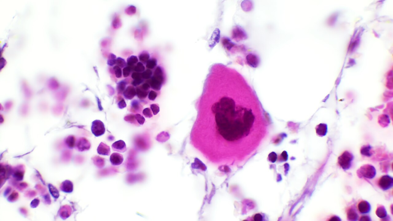
Megakaryocyte in the red bone marrow.
Summary
edit| Description |
Українська: Мікрофотографія зрізу епіфіза трубчастої кістки лабораторного щура, на якій видно найбільшу клітину червоного кісткового мозку – мегакаріоцит. Розмір клітини 100 мкм. Клітина має велике ядро і поліплоїдний набір хромосом. Цитоплазматичні відростки мегакаріоцитів проникають в ендотеліальні клітини в капілярах червоного кісткового мозку, а потім відокремлюються, утворюючи тромбоцити. Товщина зрізу 7 мкм. Забарвлення гематоксиліном і еозином. Сфотографовано на мікроскопі ZEISS Primo Star 3 за допомогою кольорової цифрової камери Axiocam 208 і програми Zen (ZEISS). Збільшення — 1000 разів.
English: A photomicrograph of a section of the epiphysis of a tubular bone of a laboratory rat, which shows the largest red bone marrow cell - a megakaryocyte. The size of the cell is 100 μm. The cell has a large nucleus and a polyploid set of chromosomes. The cytoplasmic processes of the megakaryocyte penetrate endothelial cells in the red bone marrow capillaries and then separate to form platelets. The thickness of the section is 7 µm. Staining with hematoxylin and eosin. Photographed on a ZEISS Primo Star 3 microscope using an Axiocam 208 color digital camera and Zen software (ZEISS).1000x magnification. |
| Date | |
| Source | Own work |
| Author | Yarolav Andriichuk Alina Golas Oksana Korotka |
Licensing
editI, the copyright holder of this work, hereby publish it under the following license:
This file is licensed under the Creative Commons Attribution-Share Alike 4.0 International license.
- You are free:
- to share – to copy, distribute and transmit the work
- to remix – to adapt the work
- Under the following conditions:
- attribution – You must give appropriate credit, provide a link to the license, and indicate if changes were made. You may do so in any reasonable manner, but not in any way that suggests the licensor endorses you or your use.
- share alike – If you remix, transform, or build upon the material, you must distribute your contributions under the same or compatible license as the original.
| This image was uploaded as part of Science Photo Competition 2023 in Ukraine. |
File history
Click on a date/time to view the file as it appeared at that time.
| Date/Time | Thumbnail | Dimensions | User | Comment | |
|---|---|---|---|---|---|
| current | 18:37, 20 December 2023 |  | 3,840 × 2,160 (5.08 MB) | Yarolav Andriichuk (talk | contribs) | Uploaded own work with UploadWizard |
You cannot overwrite this file.
File usage on Commons
There are no pages that use this file.