File:2011 Autophagy.tif

Size of this JPG preview of this TIF file: 439 × 600 pixels. Other resolutions: 176 × 240 pixels | 351 × 480 pixels | 562 × 768 pixels | 749 × 1,024 pixels | 1,499 × 2,048 pixels | 2,609 × 3,565 pixels.
Original file (2,609 × 3,565 pixels, file size: 26.65 MB, MIME type: image/tiff)
Captions
Captions
Add a one-line explanation of what this file represents
Summary edit
| Description |
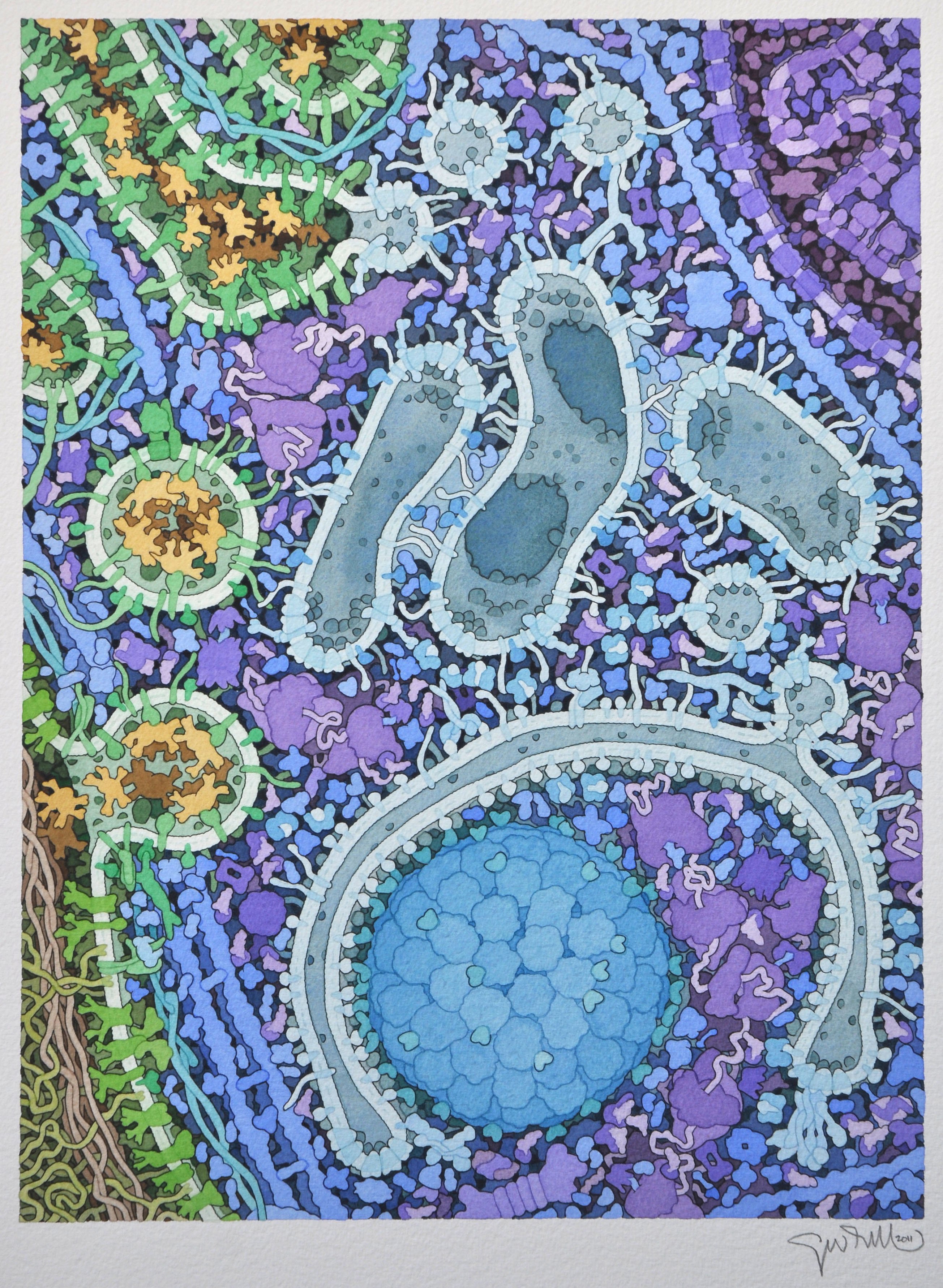
English: The formation of an autophagosome is shown, with Golgi at top left, a mitochondrion at top right, and the autophagosome at bottom center. This illustration was created in collaboration with Daniel Klionsky at the University of Michigan as a cover for the journal Cell.
For more information, see the Molecule of the Month feature on Aminopeptidase 1 and Autophagy. |
| Date | |
| Source | https://pdb101.rcsb.org/sci-art/goodsell-gallery/autophagy |
| Author | David Goodsell |
Licensing edit
This file is licensed under the Creative Commons Attribution 4.0 International license.
- You are free:
- to share – to copy, distribute and transmit the work
- to remix – to adapt the work
- Under the following conditions:
- attribution – You must give appropriate credit, provide a link to the license, and indicate if changes were made. You may do so in any reasonable manner, but not in any way that suggests the licensor endorses you or your use.
File history
Click on a date/time to view the file as it appeared at that time.
| Date/Time | Thumbnail | Dimensions | User | Comment | |
|---|---|---|---|---|---|
| current | 06:23, 13 July 2019 |  | 2,609 × 3,565 (26.65 MB) | Evolution and evolvability (talk | contribs) | User created page with UploadWizard |
You cannot overwrite this file.
File usage on Commons
There are no pages that use this file.
File usage on other wikis
The following other wikis use this file:
- Usage on zh.wikipedia.org